• 文献检索
  • 文档翻译
  • 深度研究
  • 学术资讯
  • Suppr Zotero 插件Zotero 插件
  • 邀请有礼
  • 套餐&价格
  • 历史记录
应用&插件
Suppr Zotero 插件Zotero 插件浏览器插件Mac 客户端Windows 客户端微信小程序
定价
高级版会员购买积分包购买API积分包
服务
文献检索文档翻译深度研究API 文档MCP 服务
关于我们
关于 Suppr公司介绍联系我们用户协议隐私条款
关注我们

Suppr 超能文献

核心技术专利:CN118964589B侵权必究
粤ICP备2023148730 号-1Suppr @ 2026

文献检索

告别复杂PubMed语法,用中文像聊天一样搜索,搜遍4000万医学文献。AI智能推荐,让科研检索更轻松。

立即免费搜索

文件翻译

保留排版,准确专业,支持PDF/Word/PPT等文件格式,支持 12+语言互译。

免费翻译文档

深度研究

AI帮你快速写综述,25分钟生成高质量综述,智能提取关键信息,辅助科研写作。

立即免费体验

使用原子力显微镜对内向整流型ATP敏感性钾离子通道蛋白ROMK1进行成像。

Imaging ROMK1 inwardly rectifying ATP-sensitive K+ channel protein using atomic force microscopy.

作者信息

Henderson R M, Schneider S, Li Q, Hornby D, White S J, Oberleithner H

机构信息

Department of Pharmacology, University of Cambridge, United Kingdom.

出版信息

Proc Natl Acad Sci U S A. 1996 Aug 6;93(16):8756-60. doi: 10.1073/pnas.93.16.8756.

DOI:10.1073/pnas.93.16.8756
PMID:8710944
原文链接:https://pmc.ncbi.nlm.nih.gov/articles/PMC38746/
Abstract

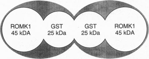
The inwardly rectifying K+ channel ROMK1 has been implicated as being significant in K+ secretion in the distal nephron. ROMK1 has been shown by immunocytochemistry to be expressed in relevant nephron segments. The development of the atomic force microscope has made possible the production of high resolution images of small particles, including a variety of biological macromolecules. Recently, a fusion protein of glutathione S-transferase (GST) and ROMK1 (ROMK1-GST) has been used to produce a polyclonal antibody for immunolocalization of ROMK1. We have used atomic force microscopy to examine ROMK1-GST and the native ROMK1 polypeptide cleaved from GST. Imaging was conducted with the proteins in physiological solutions attached to mica. ROMK1-GST appears in images as a particle composed of two units of similar size. Analyses of images indicate that the two units have volumes of approximately 118 nm3, which is close to the theoretical volume of a globular protein of approximately 65 kDa (the molecular mass of ROMK1-GST). Native GST exists as a dimer, and the images obtained here are consistent with the ROMK1-GST fusion protein's existence as a heterodimer. In experiments on ROMK1 in aqueous solution, single molecules appear to aggregate, but contact to the mica was maintained. Addition of ATP to the solution produced a change in height of the aggregates. This change (which was reversible) suggests that ATP induces a structural change in the ROMK1 protein. The data show that atomic force microscopy is a useful tool for examination of purified protein molecules under near-physiological conditions, and furthermore, that structural alterations in the proteins may be continuously investigated.

摘要

内向整流钾通道ROMK1被认为在远端肾单位的钾分泌中起重要作用。免疫细胞化学显示ROMK1在相关肾单位节段中表达。原子力显微镜的发展使得生成包括各种生物大分子在内的小颗粒的高分辨率图像成为可能。最近,谷胱甘肽S-转移酶(GST)与ROMK1的融合蛋白(ROMK1-GST)已被用于制备用于ROMK1免疫定位的多克隆抗体。我们使用原子力显微镜检查ROMK1-GST和从GST切割下来的天然ROMK1多肽。在附着于云母的生理溶液中对蛋白质进行成像。ROMK1-GST在图像中表现为一个由两个大小相似的单元组成的颗粒。图像分析表明,这两个单元的体积约为118 nm³,接近约65 kDa(ROMK1-GST的分子量)球形蛋白的理论体积。天然GST以二聚体形式存在,此处获得的图像与ROMK1-GST融合蛋白作为异二聚体的存在一致。在水溶液中对ROMK1进行的实验中,单分子似乎会聚集,但与云母的接触得以维持。向溶液中添加ATP会使聚集体的高度发生变化。这种变化(是可逆的)表明ATP诱导了ROMK1蛋白的结构变化。数据表明,原子力显微镜是在接近生理条件下检查纯化蛋白质分子的有用工具,此外,还可以持续研究蛋白质中的结构改变。

https://cdn.ncbi.nlm.nih.gov/pmc/blobs/bb9b/38746/7c64c53c5ce3/pnas01520-0614-c.jpg
https://cdn.ncbi.nlm.nih.gov/pmc/blobs/bb9b/38746/d7893d6af1a9/pnas01520-0613-a.jpg
https://cdn.ncbi.nlm.nih.gov/pmc/blobs/bb9b/38746/95c8c49c7c4b/pnas01520-0614-a.jpg
https://cdn.ncbi.nlm.nih.gov/pmc/blobs/bb9b/38746/4e265f860316/pnas01520-0614-b.jpg
https://cdn.ncbi.nlm.nih.gov/pmc/blobs/bb9b/38746/7c64c53c5ce3/pnas01520-0614-c.jpg

相似文献

1
Imaging ROMK1 inwardly rectifying ATP-sensitive K+ channel protein using atomic force microscopy.使用原子力显微镜对内向整流型ATP敏感性钾离子通道蛋白ROMK1进行成像。
Proc Natl Acad Sci U S A. 1996 Aug 6;93(16):8756-60. doi: 10.1073/pnas.93.16.8756.
2
Structural activity of a cloned potassium channel (ROMK1) monitored with the atomic force microscope: the "molecular-sandwich" technique.用原子力显微镜监测克隆钾通道(ROMK1)的结构活性:“分子三明治”技术。
Proc Natl Acad Sci U S A. 1997 Dec 9;94(25):14144-9. doi: 10.1073/pnas.94.25.14144.
3
Molecular weights of individual proteins correlate with molecular volumes measured by atomic force microscopy.
Pflugers Arch. 1998 Feb;435(3):362-7. doi: 10.1007/s004240050524.
4
Dissimilarity in the channel intrinsic stability among the bacterial KcsA and the inwardly rectifying potassium channel ROMK1.细菌 KcsA 和内向整流钾通道 ROMK1 的通道本征稳定性存在差异。
Biochimie. 2009 Nov-Dec;91(11-12):1426-33. doi: 10.1016/j.biochi.2009.08.003. Epub 2009 Aug 12.
5
Regulation of ROMK1 K+ channel activity involves phosphorylation processes.ROMK1钾离子通道活性的调节涉及磷酸化过程。
Proc Natl Acad Sci U S A. 1994 Aug 16;91(17):8077-81. doi: 10.1073/pnas.91.17.8077.
6
Gating of inwardly rectifying K+ channels localized to a single negatively charged residue.内向整流钾通道的门控定位在单个带负电荷的残基上。
Nature. 1994 Sep 15;371(6494):246-9. doi: 10.1038/371246a0.
7
Regulation of ROMK1 channel by protein kinase A via a phosphatidylinositol 4,5-bisphosphate-dependent mechanism.蛋白激酶A通过磷脂酰肌醇4,5-二磷酸依赖性机制对ROMK1通道进行调控。
Proc Natl Acad Sci U S A. 1999 May 11;96(10):5820-5. doi: 10.1073/pnas.96.10.5820.
8
Tetanus toxin abolishes exocytosis of ROMK1 induced by inhibition of protein tyrosine kinase.破伤风毒素可消除因蛋白酪氨酸激酶抑制而诱导的ROMK1胞吐作用。
Am J Physiol Renal Physiol. 2003 Mar;284(3):F510-7. doi: 10.1152/ajprenal.00309.2002. Epub 2002 Dec 30.
9
Alternative splicing of human inwardly rectifying K+ channel ROMK1 mRNA.人类内向整流钾通道ROMK1 mRNA的可变剪接
Mol Pharmacol. 1994 May;45(5):854-60.
10
Functional and structural characterization of PKA-mediated pHi gating of ROMK1 channels.蛋白激酶A介导的ROMK1通道pH值门控的功能与结构特征
J Mol Graph Model. 2008 Oct;27(3):332-41. doi: 10.1016/j.jmgm.2008.06.001. Epub 2008 Jun 8.

引用本文的文献

1
Revealing the ultrastructure of the membrane pores of intact cells by atomic force microscopy.用原子力显微镜揭示完整细胞的膜孔超微结构。
Heliyon. 2019 Oct 15;5(10):e02636. doi: 10.1016/j.heliyon.2019.e02636. eCollection 2019 Oct.
2
The Enzymatic Activity of APOBE3G Multimers.APOBE3G 多聚体的酶活性。
Sci Rep. 2018 Dec 18;8(1):17953. doi: 10.1038/s41598-018-36372-6.
3
Partially Assembled Nucleosome Structures at Atomic Detail.原子水平细节的部分组装核小体结构

本文引用的文献

1
Atomic force microscope.原子力显微镜
Phys Rev Lett. 1986 Mar 3;56(9):930-933. doi: 10.1103/PhysRevLett.56.930.
2
Renal potassium channels: an overview.肾钾通道:概述
Kidney Int. 1995 Oct;48(4):1004-9. doi: 10.1038/ki.1995.382.
3
Biological applications of atomic force microscopy.原子力显微镜的生物学应用。
Biophys J. 2017 Feb 7;112(3):460-472. doi: 10.1016/j.bpj.2016.10.041. Epub 2016 Dec 28.
4
Specific transfection of inflamed brain by macrophages: a new therapeutic strategy for neurodegenerative diseases.通过巨噬细胞对炎症大脑的特异性转染:治疗神经退行性疾病的新策略。
PLoS One. 2013 Apr 19;8(4):e61852. doi: 10.1371/journal.pone.0061852. Print 2013.
5
Using DNA as a fiducial marker to study SMC complex interactions with the atomic force microscope.利用 DNA 作为示踪物,用原子力显微镜研究 SMC 复合物与原子力显微镜的相互作用。
Biophys J. 2012 Feb 22;102(4):839-48. doi: 10.1016/j.bpj.2012.01.022. Epub 2012 Feb 21.
6
Polyelectrolyte complex optimization for macrophage delivery of redox enzyme nanoparticles.用于向巨噬细胞递送氧化还原酶纳米颗粒的聚电解质复合物优化。
Nanomedicine (Lond). 2011 Jan;6(1):25-42. doi: 10.2217/nnm.10.129.
7
Single protein molecule mapping with magnetic atomic force microscopy.利用磁原子力显微镜对单个蛋白质分子进行成像。
Biophys J. 2010 Feb 3;98(3):478-87. doi: 10.1016/j.bpj.2009.10.021.
8
Single-molecule dynamics of the DNA-EcoRII protein complexes revealed with high-speed atomic force microscopy.利用高速原子力显微镜揭示的DNA-EcoRII蛋白复合物的单分子动力学
Biochemistry. 2009 Nov 10;48(44):10492-8. doi: 10.1021/bi9010368.
9
Molecular mechanism underlying RAG1/RAG2 synaptic complex formation.RAG1/RAG2 突触复合体形成的分子机制。
J Biol Chem. 2009 Jul 31;284(31):20956-65. doi: 10.1074/jbc.M109.028977. Epub 2009 Jun 5.
10
Probing cellular microenvironments and tissue remodeling by atomic force microscopy.利用原子力显微镜探测细胞微环境和组织重塑
Pflugers Arch. 2008 Apr;456(1):29-49. doi: 10.1007/s00424-007-0398-9. Epub 2007 Dec 6.
Am J Physiol. 1994 Jan;266(1 Pt 1):C1-21. doi: 10.1152/ajpcell.1994.266.1.C1.
4
Promises and problems of biological atomic force microscopy.
J Microsc. 1993 Sep;171(Pt 3):183-98. doi: 10.1111/j.1365-2818.1993.tb03375.x.
5
Primary structure and functional properties of an epithelial K channel.一种上皮钾通道的一级结构和功能特性
Am J Physiol. 1994 Mar;266(3 Pt 1):C809-24. doi: 10.1152/ajpcell.1994.266.3.C809.
6
Regulation of ROMK1 K+ channel activity involves phosphorylation processes.ROMK1钾离子通道活性的调节涉及磷酸化过程。
Proc Natl Acad Sci U S A. 1994 Aug 16;91(17):8077-81. doi: 10.1073/pnas.91.17.8077.
7
Biological applications of scanning probe microscopies.扫描探针显微镜的生物学应用。
Prog Biophys Mol Biol. 1994;61(2):131-85. doi: 10.1016/0079-6107(94)90008-6.
8
Probing chromatin with the scanning force microscope.用扫描力显微镜探测染色质。
Chromosoma. 1994 Jul;103(4):231-6. doi: 10.1007/BF00352247.
9
The scanning force microscopy of DNA in air and in n-propanol using new spreading agents.使用新型铺展剂在空气和正丙醇中对DNA进行扫描力显微镜观察。
FEBS Lett. 1994 Nov 21;355(1):91-5. doi: 10.1016/0014-5793(94)01166-4.
10
Biomolecular imaging with the atomic force microscope.利用原子力显微镜进行生物分子成像。
Annu Rev Biophys Biomol Struct. 1994;23:115-39. doi: 10.1146/annurev.bb.23.060194.000555.