Buchs P A, Muller D
Pharmacology, Centre Médical Universitaire, Geneva, Switzerland.
Proc Natl Acad Sci U S A. 1996 Jul 23;93(15):8040-5. doi: 10.1073/pnas.93.15.8040.
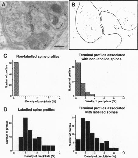
Long-term potentiation (LTP), an increase in synaptic efficacy believed to underlie learning and memory mechanisms, has been proposed to involve structural modifications of synapses. Precise identification of the morphological changes associated with LTP has however been hindered by the difficulty in distinguishing potentiated or activated from nonstimulated synapses. Here we used a cytochemical method that allowed detection in CA1 hippocampus at the electron microscopy level of a stimulation-specific, D-AP5-sensitive accumulation of calcium in postsynaptic spines and presynaptic terminals following application of high-frequency trains. Morphometric analyses carried out 30-40 min after LTP induction revealed dramatic ultrastructural differences between labeled and nonlabeled synapses. The majority of labeled synapses (60%) exhibited perforated postsynaptic densities, whereas this proportion was only 20% in nonlabeled synaptic contacts. Labeled synaptic profiles were also characterized by a larger apposition zone between pre- and postsynaptic structures, longer postsynaptic densities, and enlarged spine profiles. These results add strong support to the idea that ultrastructural modifications and specifically an increase in perforated synapses are associated with LTP induction in field CA1 of hippocampus and they suggest that a majority of activated contacts may exhibit such changes.
长期增强作用(LTP)是一种突触效能的增强,被认为是学习和记忆机制的基础,有人提出它涉及突触的结构修饰。然而,由于难以区分增强或激活的突触与未受刺激的突触,精确识别与LTP相关的形态学变化受到了阻碍。在这里,我们使用了一种细胞化学方法,该方法能够在电子显微镜水平上检测到,在海马体CA1区,高频串刺激后,突触后棘突和突触前终末中出现的一种刺激特异性、对D-AP5敏感的钙积累。在LTP诱导后30 - 40分钟进行的形态计量分析显示,标记突触和未标记突触之间存在显著的超微结构差异。大多数标记突触(60%)表现出穿孔的突触后致密物,而在未标记的突触接触中这一比例仅为20%。标记的突触轮廓还具有突触前和突触后结构之间更大的并置区、更长的突触后致密物以及扩大的棘突轮廓等特征。这些结果为超微结构修饰,特别是穿孔突触的增加与海马体CA1区LTP诱导相关这一观点提供了有力支持,并且表明大多数激活的突触接触可能会出现这种变化。