• 文献检索
  • 文档翻译
  • 深度研究
  • 学术资讯
  • Suppr Zotero 插件Zotero 插件
  • 邀请有礼
  • 套餐&价格
  • 历史记录
应用&插件
Suppr Zotero 插件Zotero 插件浏览器插件Mac 客户端Windows 客户端微信小程序
定价
高级版会员购买积分包购买API积分包
服务
文献检索文档翻译深度研究API 文档MCP 服务
关于我们
关于 Suppr公司介绍联系我们用户协议隐私条款
关注我们

Suppr 超能文献

核心技术专利:CN118964589B侵权必究
粤ICP备2023148730 号-1Suppr @ 2026

文献检索

告别复杂PubMed语法,用中文像聊天一样搜索,搜遍4000万医学文献。AI智能推荐,让科研检索更轻松。

立即免费搜索

文件翻译

保留排版,准确专业,支持PDF/Word/PPT等文件格式,支持 12+语言互译。

免费翻译文档

深度研究

AI帮你快速写综述,25分钟生成高质量综述,智能提取关键信息,辅助科研写作。

立即免费体验

单独的钙并不能完全激活细肌丝,以使肌球蛋白头部(S1)与僵直肌原纤维结合。

Calcium alone does not fully activate the thin filament for S1 binding to rigor myofibrils.

作者信息

Swartz D R, Moss R L, Greaser M L

机构信息

Department of Anatomy, Indiana University Medical School, Indianapolis 46202, USA.

出版信息

Biophys J. 1996 Oct;71(4):1891-904. doi: 10.1016/S0006-3495(96)79388-8.

DOI:10.1016/S0006-3495(96)79388-8
PMID:8889164
原文链接:https://pmc.ncbi.nlm.nih.gov/articles/PMC1233656/
Abstract

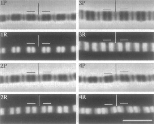
Skeletal muscle contraction is regulated by calcium via troponin and tropomyosin and appears to involve cooperative activation of cross-bridge binding to actin. We studied the regulation of fluorescent myosin subfragment 1 (fS1) binding to rigor myofibrils over a wide range of fS1 and calcium levels using highly sensitive imaging techniques. At low calcium and low fS1, the fluorescence was restricted to the actin-myosin overlap region. At high calcium and very low fS1, the fluorescence was still predominantly in the overlap region. The ratio of nonoverlap to overlap fluorescence intensity showed that increases in the fS1 level resulted in a shift in maximum fluorescence from the overlap to the nonoverlap region at both low and high calcium; this transition occurred at lower fS1 levels in myofibrils with high calcium. At a fixed fS1 level, increases in calcium also resulted in a shift in maximum fluorescence from the overlap region to the nonoverlap region. These results suggest that calcium alone does not fully activate the thin filament for rigor S1 binding and that, even at high calcium, the thin filament is not activated along its entire length.

摘要

骨骼肌收缩由钙通过肌钙蛋白和原肌球蛋白进行调节,并且似乎涉及横桥与肌动蛋白结合的协同激活。我们使用高灵敏度成像技术,在很宽的肌球蛋白亚片段1(fS1)和钙水平范围内,研究了荧光fS1与僵直肌原纤维结合的调节。在低钙和低fS1条件下,荧光局限于肌动蛋白 - 肌球蛋白重叠区域。在高钙和极低fS1条件下,荧光仍主要位于重叠区域。非重叠与重叠荧光强度的比值表明,fS1水平的增加导致在低钙和高钙条件下,最大荧光从重叠区域向非重叠区域偏移;在高钙的肌原纤维中,这种转变发生在较低的fS1水平。在固定的fS1水平下,钙的增加也导致最大荧光从重叠区域向非重叠区域偏移。这些结果表明,仅钙本身并不能完全激活细肌丝以实现与僵直S1的结合,并且即使在高钙条件下,细肌丝也并非沿其全长被激活。

https://cdn.ncbi.nlm.nih.gov/pmc/blobs/66ab/1233656/7db30ef3112d/biophysj00044-0244-a.jpg
https://cdn.ncbi.nlm.nih.gov/pmc/blobs/66ab/1233656/2b26bf027574/biophysj00044-0239-a.jpg
https://cdn.ncbi.nlm.nih.gov/pmc/blobs/66ab/1233656/72eaf5c5e174/biophysj00044-0239-b.jpg
https://cdn.ncbi.nlm.nih.gov/pmc/blobs/66ab/1233656/e17dec2810c3/biophysj00044-0240-a.jpg
https://cdn.ncbi.nlm.nih.gov/pmc/blobs/66ab/1233656/bbc044d0ea56/biophysj00044-0240-b.jpg
https://cdn.ncbi.nlm.nih.gov/pmc/blobs/66ab/1233656/3cd6a10be0fe/biophysj00044-0241-a.jpg
https://cdn.ncbi.nlm.nih.gov/pmc/blobs/66ab/1233656/5931b7cfa55c/biophysj00044-0243-a.jpg
https://cdn.ncbi.nlm.nih.gov/pmc/blobs/66ab/1233656/7db30ef3112d/biophysj00044-0244-a.jpg

相似文献

1
Calcium alone does not fully activate the thin filament for S1 binding to rigor myofibrils.单独的钙并不能完全激活细肌丝,以使肌球蛋白头部(S1)与僵直肌原纤维结合。
Biophys J. 1996 Oct;71(4):1891-904. doi: 10.1016/S0006-3495(96)79388-8.
2
Influence of ADP on cross-bridge-dependent activation of myofibrillar thin filaments.二磷酸腺苷对肌原纤维细肌丝横桥依赖性激活的影响。
Biophys J. 2000 Jun;78(6):3103-11. doi: 10.1016/S0006-3495(00)76847-0.
3
Characteristics of troponin C binding to the myofibrillar thin filament: extraction of troponin C is not random along the length of the thin filament.肌钙蛋白C与肌原纤维细肌丝结合的特性:肌钙蛋白C沿细肌丝长度的提取并非随机的。
Biophys J. 1997 Jul;73(1):293-305. doi: 10.1016/S0006-3495(97)78070-6.
4
Regulation of binding of subfragment 1 in isolated rigor myofibrils.分离的强直肌原纤维中肌球蛋白亚片段1结合的调节
J Cell Biol. 1990 Dec;111(6 Pt 2):2989-3001. doi: 10.1083/jcb.111.6.2989.
5
Myofibrillar troponin exists in three states and there is signal transduction along skeletal myofibrillar thin filaments.肌原纤维肌钙蛋白以三种状态存在,并且沿着骨骼肌原纤维细肌丝存在信号转导。
J Mol Biol. 2006 Aug 18;361(3):420-35. doi: 10.1016/j.jmb.2006.05.078. Epub 2006 Jun 30.
6
Differences between cardiac and skeletal troponin interaction with the thin filament probed by troponin exchange in skeletal myofibrils.通过肌钙蛋白交换探测骨骼肌肌原纤维中肌钙蛋白与细肌丝相互作用时心肌与骨骼肌肌钙蛋白的差异。
Biophys J. 2009 Jul 8;97(1):183-94. doi: 10.1016/j.bpj.2009.04.023.
7
Rigor cross-bridges bind to two actin monomers in thin filaments of rabbit psoas muscle.强直横桥与兔腰大肌细肌丝中的两个肌动蛋白单体结合。
J Mol Biol. 1995 Apr 28;248(2):294-307.
8
Structural studies of rigor bovine myofibrils using fluorescence microscopy. II. Influence of sarcomere length on the binding of myosin subfragment-1, alpha-actinin and G-actin to rigor myofibrils.利用荧光显微镜对僵直牛肌原纤维进行的结构研究。II. 肌节长度对肌球蛋白亚片段-1、α-辅肌动蛋白和G-肌动蛋白与僵直肌原纤维结合的影响。
Meat Sci. 1993;33(2):157-90. doi: 10.1016/0309-1740(93)90058-P.
9
Detection of fluorescently labeled actin-bound cross-bridges in actively contracting myofibrils.在活跃收缩的肌原纤维中检测荧光标记的肌动蛋白结合横桥。
Biophys J. 2000 Mar;78(3):1449-57. doi: 10.1016/S0006-3495(00)76698-7.
10
Actin-tropomyosin activation of myosin subfragment 1 ATPase and thin filament cooperativity. The role of tropomyosin flexibility and end-to-end interactions.肌动蛋白-原肌球蛋白对肌球蛋白亚片段1 ATP酶的激活及细肌丝协同性。原肌球蛋白柔韧性及端对端相互作用的作用。
Biochemistry. 1997 Nov 4;36(44):13449-54. doi: 10.1021/bi971568w.

引用本文的文献

1
Troponin Structural Dynamics in the Native Cardiac Thin Filament Revealed by Cryo Electron Microscopy.冷冻电子显微镜揭示天然心脏细肌丝中的肌钙蛋白结构动力学
J Mol Biol. 2024 Mar 15;436(6):168498. doi: 10.1016/j.jmb.2024.168498. Epub 2024 Feb 20.
2
Mouse Models of Cardiomyopathies Caused by Mutations in Troponin C.肌钙蛋白 C 突变所致心肌病的小鼠模型。
Int J Mol Sci. 2023 Aug 2;24(15):12349. doi: 10.3390/ijms241512349.
3
Cryo-Electron Microscopy Reveals Cardiac Myosin Binding Protein-C M-Domain Interactions with the Thin Filament.

本文引用的文献

1
Determination of serum proteins by means of the biuret reaction.通过双缩脲反应测定血清蛋白。
J Biol Chem. 1949 Feb;177(2):751-66.
2
Theoretical model for the cooperative equilibrium binding of myosin subfragment 1 to the actin-troponin-tropomyosin complex.肌球蛋白亚片段1与肌动蛋白-肌钙蛋白-原肌球蛋白复合物协同平衡结合的理论模型。
Proc Natl Acad Sci U S A. 1980 Jun;77(6):3186-90. doi: 10.1073/pnas.77.6.3186.
3
Subsarcomeric distribution of calcium in demembranated fibers of rabbit psoas muscle.兔腰大肌去膜纤维中钙的肌小节下分布
冷冻电镜揭示了肌球蛋白结合蛋白-C M 结构域与细肌丝的相互作用。
J Mol Biol. 2022 Dec 30;434(24):167879. doi: 10.1016/j.jmb.2022.167879. Epub 2022 Nov 10.
4
cMyBP-C phosphorylation modulates the time-dependent slowing of unloaded shortening in murine skinned myocardium.cMyBP-C 磷酸化调节去负荷缩短时心肌肌球蛋白速度的时相依赖性减慢。
J Gen Physiol. 2021 Mar 1;153(3). doi: 10.1085/jgp.202012782.
5
Fission yeast tropomyosin specifies directed transport of myosin-V along actin cables.裂殖酵母原肌球蛋白沿肌动蛋白缆定向运输肌球蛋白-V。
Mol Biol Cell. 2014 Jan;25(1):66-75. doi: 10.1091/mbc.E13-04-0200. Epub 2013 Nov 6.
6
The myosin duty ratio tunes the calcium sensitivity and cooperative activation of the thin filament.肌球蛋白做功比调节细肌丝的钙敏感性和协同激活。
Biochemistry. 2013 Sep 17;52(37):6437-44. doi: 10.1021/bi400262h. Epub 2013 Sep 3.
7
Regulation of fibre contraction in a rat model of myocardial ischemia.心肌缺血大鼠模型中纤维收缩的调节。
PLoS One. 2010 Mar 4;5(3):e9528. doi: 10.1371/journal.pone.0009528.
8
Tropomyosin and myosin-II cellular levels promote actomyosin ring assembly in fission yeast.原肌球蛋白和肌球蛋白-II 的细胞水平促进有丝分裂酵母中肌动球蛋白环的组装。
Mol Biol Cell. 2010 Mar 15;21(6):989-1000. doi: 10.1091/mbc.e09-10-0852. Epub 2010 Jan 28.
9
Differences between cardiac and skeletal troponin interaction with the thin filament probed by troponin exchange in skeletal myofibrils.通过肌钙蛋白交换探测骨骼肌肌原纤维中肌钙蛋白与细肌丝相互作用时心肌与骨骼肌肌钙蛋白的差异。
Biophys J. 2009 Jul 8;97(1):183-94. doi: 10.1016/j.bpj.2009.04.023.
10
Phosphorylation of tropomyosin extends cooperative binding of myosin beyond a single regulatory unit.原肌球蛋白的磷酸化将肌球蛋白的协同结合扩展到单个调节单位之外。
Cell Motil Cytoskeleton. 2009 Jan;66(1):10-23. doi: 10.1002/cm.20321.
Biophys J. 1993 Jan;64(1):211-22. doi: 10.1016/S0006-3495(93)81358-4.
4
Calcium ions and the structure of muscle actin filament. An X-ray diffraction study.钙离子与肌肉肌动蛋白丝的结构。一项X射线衍射研究。
J Mol Biol. 1993 Jan 20;229(2):279-85. doi: 10.1006/jmbi.1993.1032.
5
Regulation of the interaction between actin and myosin subfragment 1: evidence for three states of the thin filament.肌动蛋白与肌球蛋白亚片段1之间相互作用的调节:细肌丝三种状态的证据。
Biophys J. 1993 Aug;65(2):693-701. doi: 10.1016/S0006-3495(93)81110-X.
6
The three-dimensional structure of a molecular motor.分子马达的三维结构。
Trends Biochem Sci. 1994 Mar;19(3):129-34. doi: 10.1016/0968-0004(94)90206-2.
7
Ca(2+)-induced tropomyosin movement in Limulus thin filaments revealed by three-dimensional reconstruction.通过三维重建揭示鲎细肌丝中钙离子诱导的原肌球蛋白运动。
Nature. 1994 Mar 3;368(6466):65-7. doi: 10.1038/368065a0.
8
The regulatory switch of the muscle thin filament: Ca2+ or myosin heads?肌肉细肌丝的调节开关:钙离子还是肌球蛋白头部?
J Muscle Res Cell Motil. 1994 Jun;15(3):232-6. doi: 10.1007/BF00123476.
9
The actomyosin interaction--shedding light on structural events: 'Plus ça change, plus c'est la même chose'.肌动球蛋白相互作用——揭示结构事件:“万变不离其宗” 。
J Muscle Res Cell Motil. 1994 Jun;15(3):227-31. doi: 10.1007/BF00123475.
10
Dynamics of the muscle thin filament regulatory switch: the size of the cooperative unit.肌肉细肌丝调节开关的动力学:协同单位的大小。
Biophys J. 1994 Jul;67(1):273-82. doi: 10.1016/S0006-3495(94)80478-3.