Wiemer E A, Wenzel T, Deerinck T J, Ellisman M H, Subramani S
Department of Biology, University of California at San Diego, La Jolla 92093-0322, USA.
J Cell Biol. 1997 Jan 13;136(1):71-80. doi: 10.1083/jcb.136.1.71.
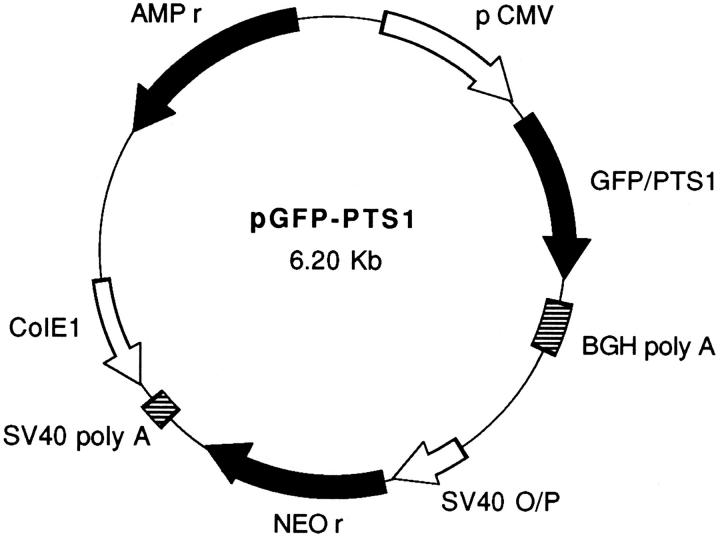
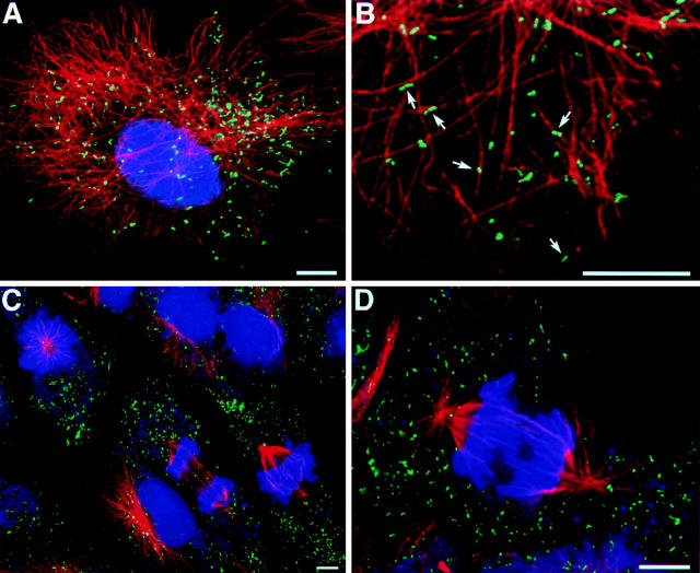
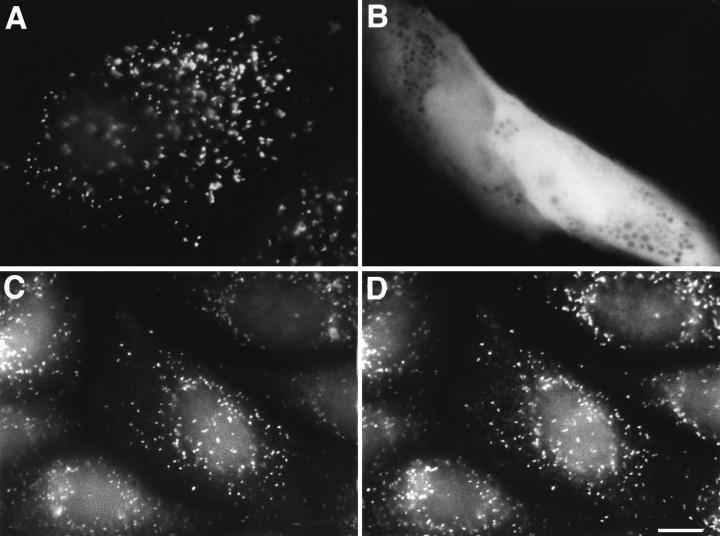
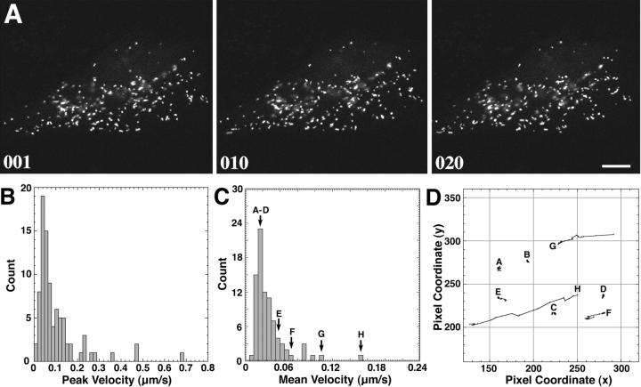
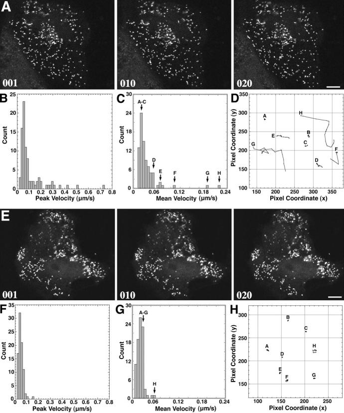
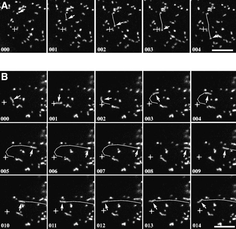
Peroxisomes in living CV1 cells were visualized by targeting the green fluorescent protein (GFP) to this subcellular compartment through the addition of a COOH-terminal peroxisomal targeting signal 1 (GFP-PTS1). The organelle dynamics were examined and analyzed using time-lapse confocal laser scanning microscopy. Two types of movement could be distinguished: a relatively slow, random, vibration-like movement displayed by the majority (approximately 95%) of the peroxisomes, and a saltatory, fast directional movement displayed by a small subset (approximately 5%) of the peroxisomes. In the latter instance, peak velocities up to 0.75 micron/s and sustained directional velocities up to 0.45 micron/s over 11.5 microns were recorded. Only the directional type of motion appeared to be energy dependent, whereas the vibrational movement continued even after the cells were depleted of energy. Treatment of cells, transiently expressing GFP-PTS1, with microtubule-destabilizing agents such as nocodazole, vinblastine, and demecolcine clearly altered peroxisome morphology and subcellular distribution and blocked the directional movement. In contrast, the microtubule-stabilizing compound paclitaxel, or the microfilament-destabilizing drugs cytochalasin B or D, did not exert these effects. High resolution confocal analysis of cells expressing GFP-PTS1 and stained with anti-tubulin antibodies revealed that many peroxisomes were associated with microtubules. The GFP-PTS1-labeled peroxisomes were found to distribute themselves in a stochastic, rather than ordered, manner to daughter cells at the time of mitosis.
通过添加羧基末端过氧化物酶体靶向信号1(GFP-PTS1)将绿色荧光蛋白(GFP)靶向到这个亚细胞区室,从而使活CV1细胞中的过氧化物酶体可视化。使用延时共聚焦激光扫描显微镜检查和分析细胞器动力学。可以区分出两种类型的运动:大多数(约95%)过氧化物酶体表现出的相对缓慢、随机、类似振动的运动,以及一小部分(约5%)过氧化物酶体表现出的跳跃式快速定向运动。在后一种情况下,记录到的峰值速度高达0.75微米/秒,在11.5微米的距离上持续定向速度高达0.45微米/秒。似乎只有定向运动类型依赖能量,而即使细胞能量耗尽,振动运动仍会继续。用微管破坏剂如诺考达唑、长春碱和秋水仙碱处理瞬时表达GFP-PTS1的细胞,明显改变了过氧化物酶体的形态和亚细胞分布,并阻断了定向运动。相比之下,微管稳定化合物紫杉醇或微丝破坏药物细胞松弛素B或D没有产生这些影响。对表达GFP-PTS1并用抗微管蛋白抗体染色的细胞进行高分辨率共聚焦分析发现,许多过氧化物酶体与微管相关。发现在有丝分裂时,GFP-PTS1标记的过氧化物酶体以随机而非有序的方式分布到子细胞中。