Rogler L E
Marion Bession Liver Center, Department of Medicine, Albert Einstein College of Medicine, Bronx, NY 10461, USA.
Am J Pathol. 1997 Feb;150(2):591-602.
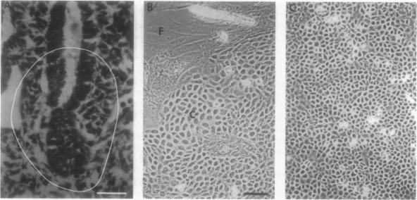
A line of hepatic endoderm cells, hepatoblast cell line 3 (HBC-3), was derived from the liver diverticulum of the mouse on day 9.5 of gestation by culture on a mitomycin C treated STON+ feeder layer in a hepatoblast culture medium consisting of Dulbecco's modified Eagle's medium, nonessential amino acids, fetal calf serum, and beta-mercaptoethanol. This line, HBC-3, stains positively for alpha-fetoprotein, albumin, and cytokeratin 14 (CK-14), protein markers expressed by the embryonic liver diverticulum, indicating that HBC-3 cells retain an undifferentiated hepatoblast phenotype. HBC-3 cells acquire hepatocyte-like ultrastructural characteristics, including bile canaliculi, peroxisomes, and glycogen granules, when maintained in culture for 3 weeks without passage. Treatment with dimethylsulfoxide or sodium butyrate induces a rapid hepatocytic differentiation. The cells cease to express alpha-fetoprotein and CK-14, maintain albumin expression, and become positive for glucose-6-phosphatase activity (a profile consistent with differentiation along the hepatocyte lineage). On Matrigel, HBC-3 cells form elaborate ductular structures, which are positive for gamma-glutamyl transpeptidase and CK-14 and CK-19 and do not express detectable amounts of albumin, a phenotypic change consistent with differentiation along the bile ductular lineage. Thus, HBC-3 cells behave in culture as bipotential hepatoblasts and provide a model system to identify factors that regulate bipotential differentiation in the liver.
一种肝内胚层细胞系,即肝母细胞系3(HBC - 3),于妊娠第9.5天从小鼠肝脏憩室中分离得到,方法是将其培养在经丝裂霉素C处理的STON +饲养层上,培养基为含有杜氏改良伊格尔培养基、非必需氨基酸、胎牛血清和β - 巯基乙醇的肝母细胞培养基。该细胞系HBC - 3对甲胎蛋白、白蛋白和细胞角蛋白14(CK - 14)呈阳性染色,这些是胚胎肝脏憩室表达的蛋白质标志物,表明HBC - 3细胞保留了未分化的肝母细胞表型。当在不传代的情况下培养3周时,HBC - 3细胞获得类似肝细胞的超微结构特征,包括胆小管、过氧化物酶体和糖原颗粒。用二甲基亚砜或丁酸钠处理可诱导快速的肝细胞分化。细胞停止表达甲胎蛋白和CK - 14,维持白蛋白表达,并对葡萄糖 - 6 - 磷酸酶活性呈阳性(这一特征与沿肝细胞谱系分化一致)。在基质胶上,HBC - 3细胞形成复杂的导管样结构,这些结构对γ - 谷氨酰转肽酶、CK - 14和CK - 19呈阳性,且不表达可检测量的白蛋白,这一表型变化与沿胆管谱系分化一致。因此,HBC - 3细胞在培养中表现为双能肝母细胞,并提供了一个模型系统来鉴定调节肝脏双能分化的因子。