Ukimura A, Deguchi H, Kitaura Y, Fujioka S, Hirasawa M, Kawamura K, Hirai K
Department of Internal Medicine, Osaka Medical College, Japan.
Am J Pathol. 1997 Jun;150(6):2061-74.
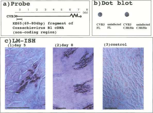
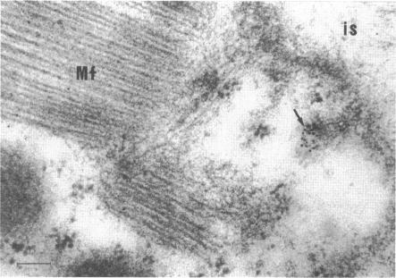
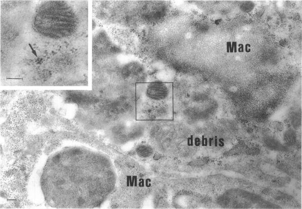
Group B Coxsackieviruses are a common cause of myocarditis. To detect the viral genome and its localization in the myocardium, we examined C3H/He mice with Coxsackievirus B3 (CVB3) myocarditis on days 5, 8, and 14 after inoculation by the reverse transcriptase polymerase chain reaction and by in situ hybridization. Sense and antisense CVB3 RNA were detected in the myocardium of all mice up to day 14 by reverse transcriptase polymerase chain reaction. Light microscopic in situ hybridization with a cDNA probe for CVB3 showed clusters of positive signals in the areas of myocardial necrosis and cell infiltration. With electron microscopic in situ hybridization, CVB3 RNA was detected in the cytoplasm of cardiocytes, between the myofibrils, near the mitochondria, and in tubular or vesicular structures. Viral RNA was also detected in necrotic debris, in the cytoplasm of macrophages, and in the cytoplasm of interstitial fibroblasts. These findings suggest that CVB3 RNA is replicated in the cytoplasm of cardiocytes, transferred into tubular or vesicular structures, released into the interstitium, and phagocytosed by macrophages. Some positive signals were also detected in the cytoplasm of cardiocytes showing close contact with infiltrating lymphocytes, suggesting that the lymphocytes recognized virus-infected cardiocytes and caused cell-mediated immune cardiocyte damage.
B组柯萨奇病毒是心肌炎的常见病因。为了检测病毒基因组及其在心肌中的定位,我们通过逆转录聚合酶链反应和原位杂交技术,对接种柯萨奇病毒B3(CVB3)后第5天、第8天和第14天的C3H/He小鼠进行了检测。通过逆转录聚合酶链反应,在所有小鼠的心肌中直至第14天都检测到了正义和反义CVB3 RNA。用CVB3的cDNA探针进行光镜原位杂交显示,在心肌坏死和细胞浸润区域有阳性信号簇。通过电镜原位杂交,在心肌细胞的细胞质中、肌原纤维之间、线粒体附近以及管状或囊泡状结构中检测到了CVB3 RNA。在坏死碎片、巨噬细胞的细胞质以及间质成纤维细胞的细胞质中也检测到了病毒RNA。这些发现表明,CVB3 RNA在心肌细胞的细胞质中复制,转移到管状或囊泡状结构中,释放到间质中,并被巨噬细胞吞噬。在与浸润淋巴细胞紧密接触的心肌细胞的细胞质中也检测到了一些阳性信号,这表明淋巴细胞识别了病毒感染的心肌细胞并导致了细胞介导的免疫性心肌细胞损伤。