Wang H L, Auerbach A, Bren N, Ohno K, Engel A G, Sine S M
Department of Physiology and Biophysics, Mayo Foundation, Rochester, Minnesota 55905, USA.
J Gen Physiol. 1997 Jun;109(6):757-66. doi: 10.1085/jgp.109.6.757.
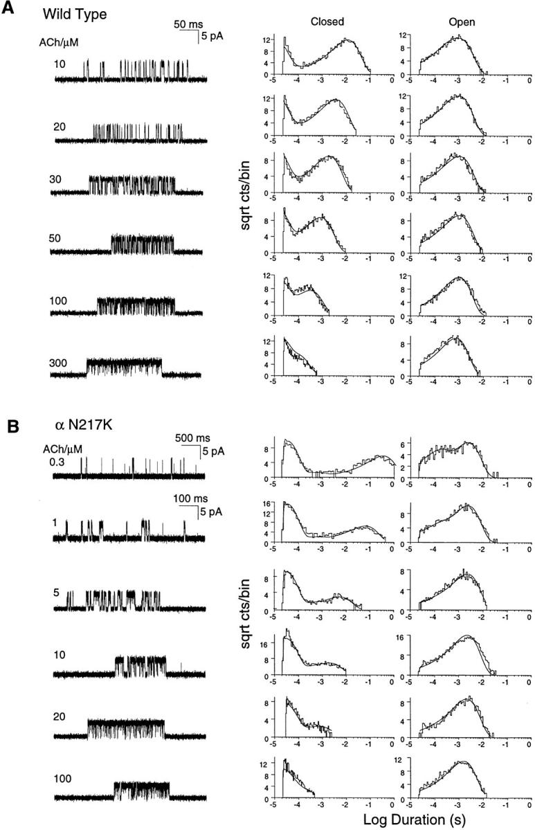
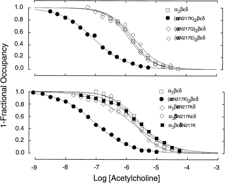
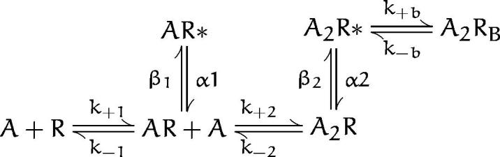
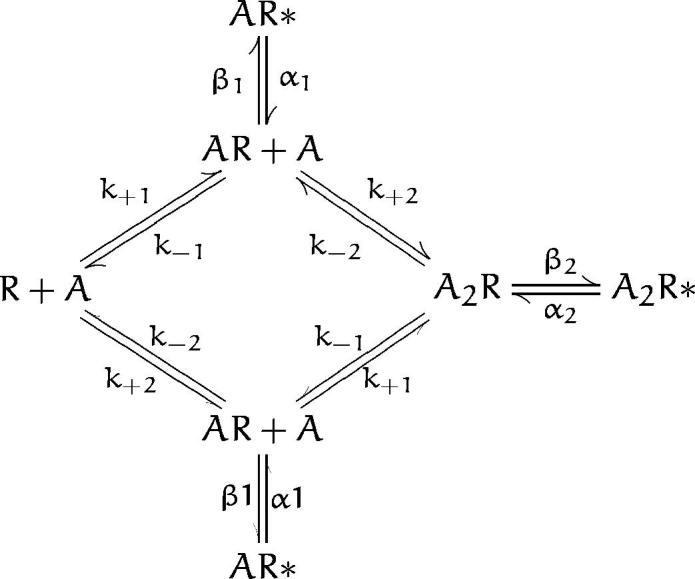
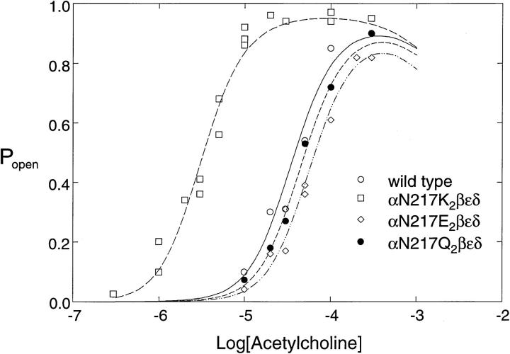
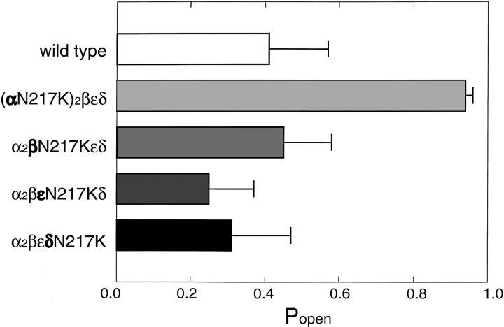
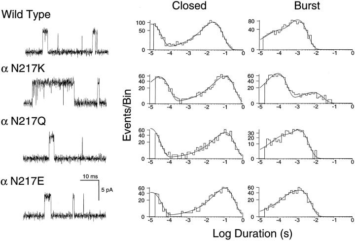
We describe the kinetic consequences of the mutation N217K in the M1 domain of the acetylcholine receptor (AChR) alpha subunit that causes a slow channel congenital myasthenic syndrome (SCCMS). We previously showed that receptors containing alpha N217K expressed in 293 HEK cells open in prolonged activation episodes strikingly similar to those observed at the SCCMS end plates. Here we use single channel kinetic analysis to show that the prolonged activation episodes result primarily from slowing of the rate of acetylcholine (ACh) dissociation from the binding site. Rate constants for channel opening and closing are also slowed but to much smaller extents. The rate constants derived from kinetic analysis also describe the concentration dependence of receptor activation, revealing a 20-fold shift in the EC50 to lower agonist concentrations for alpha N217K. The apparent affinity of ACh binding, measured by competition against the rate of 125I-alpha-bungarotoxin binding, is also enhanced 20-fold by alpha N217K. Both the slowing of ACh dissociation and enhanced apparent affinity are specific to the lysine substitution, as the glutamine and glutamate substitutions have no effect. Substituting lysine for the equivalent asparagine in the beta, epsilon, or delta subunits does not affect the kinetics of receptor activation or apparent agonist affinity. The results show that a mutation in the amino-terminal portion of the M1 domain produces a localized perturbation that stabilizes agonist bound to the resting state of the AChR.
我们描述了乙酰胆碱受体(AChR)α亚基M1结构域中N217K突变的动力学后果,该突变导致慢通道先天性肌无力综合征(SCCMS)。我们之前表明,在293 HEK细胞中表达的含有αN217K的受体在长时间激活过程中开放,这与在SCCMS终板处观察到的情况惊人地相似。在这里,我们使用单通道动力学分析表明,长时间激活过程主要是由于乙酰胆碱(ACh)从结合位点解离速率减慢所致。通道开放和关闭的速率常数也减慢,但程度要小得多。从动力学分析得出的速率常数还描述了受体激活的浓度依赖性,揭示了αN217K的EC50向较低激动剂浓度有20倍的偏移。通过与125I-α-银环蛇毒素结合速率竞争测量的ACh结合表观亲和力也因αN217K而增强了20倍。ACh解离减慢和表观亲和力增强均对赖氨酸取代具有特异性,因为谷氨酰胺和谷氨酸取代没有影响。在β、ε或δ亚基中将赖氨酸取代等效的天冬酰胺不会影响受体激活的动力学或表观激动剂亲和力。结果表明,M1结构域氨基末端部分的突变产生了局部扰动,使激动剂稳定结合于AChR的静息状态。