Caulín C, Salvesen G S, Oshima R G
The Burnham Institute (formerly the La Jolla Cancer Research Foundation), La Jolla, California 92037, USA.
J Cell Biol. 1997 Sep 22;138(6):1379-94. doi: 10.1083/jcb.138.6.1379.
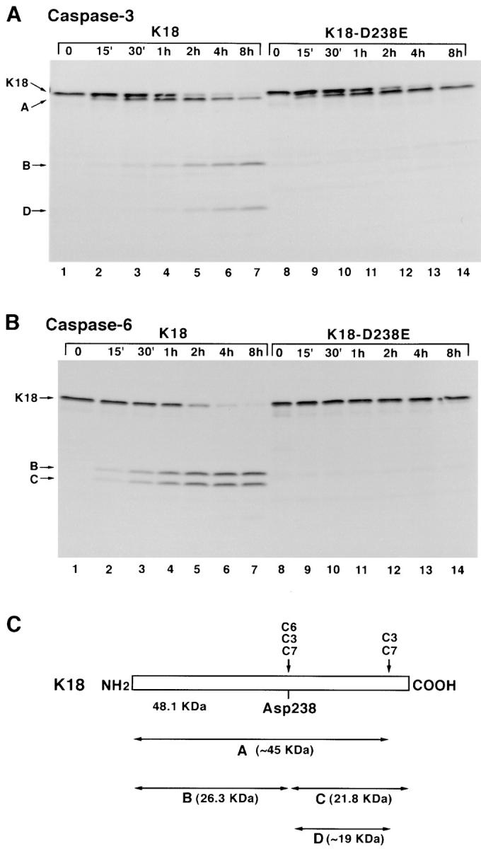
Keratins 8 (K8) and 18 (K18) are major components of intermediate filaments (IFs) of simple epithelial cells and tumors derived from such cells. Structural cell changes during apoptosis are mediated by proteases of the caspase family. During apoptosis, K18 IFs reorganize into granular structures enriched for K18 phosphorylated on serine 53. K18, but not K8, generates a proteolytic fragment during drug- and UV light-induced apoptosis; this fragment comigrates with K18 cleaved in vitro by caspase-6, -3, and -7. K18 is cleaved by caspase-6 into NH2-terminal, 26-kD and COOH-terminal, 22-kD fragments; caspase-3 and -7 additionally cleave the 22-kD fragment into a 19-kD fragment. The cleavage site common for the three caspases was the sequence VEVD/A, located in the conserved L1-2 linker region of K18. The additional site for caspases-3 and -7 that is not cleaved efficiently by caspase-6 is located in the COOH-terminal tail domain of K18. Expression of K18 with alanine instead of serine at position 53 demonstrated that cleavage during apoptosis does not require phosphorylation of serine 53. However, K18 with a glutamate instead of aspartate at position 238 was resistant to proteolysis during apoptosis. Furthermore, this cleavage site mutant appears to cause keratin filament reorganization in stably transfected clones. The identification of the L1-2 caspase cleavage site, and the conservation of the same or very similar sites in multiple other intermediate filament proteins, suggests that the processing of IFs during apoptosis may be initiated by a similar caspase cleavage.
角蛋白8(K8)和18(K18)是单层上皮细胞及由此类细胞衍生的肿瘤中间丝(IFs)的主要成分。凋亡过程中的结构细胞变化由半胱天冬酶家族的蛋白酶介导。在凋亡过程中,K18中间丝重组为富含丝氨酸53磷酸化K18的颗粒状结构。在药物和紫外线诱导的凋亡过程中,K18而非K8产生一个蛋白水解片段;该片段与体外被半胱天冬酶-6、-3和-7切割的K18共迁移。K18被半胱天冬酶-6切割成氨基末端26-kD片段和羧基末端22-kD片段;半胱天冬酶-3和-7还将22-kD片段进一步切割成19-kD片段。三种半胱天冬酶共同的切割位点是位于K18保守的L1-2连接区的序列VEVD/A。半胱天冬酶-3和-7的额外切割位点位于K18的羧基末端尾部结构域,该位点不能被半胱天冬酶-6有效切割。在53位用丙氨酸替代丝氨酸的K18表达表明,凋亡过程中的切割不需要丝氨酸53的磷酸化。然而,在238位用谷氨酸替代天冬氨酸的K18在凋亡过程中对蛋白水解具有抗性。此外,这种切割位点突变体似乎在稳定转染的克隆中导致角蛋白丝重组。L1-2半胱天冬酶切割位点的鉴定以及多个其他中间丝蛋白中相同或非常相似位点的保守性表明,凋亡过程中中间丝的加工可能由类似的半胱天冬酶切割启动。