Xiao Z, Zhang N, Murphy D B, Devreotes P N
Department of Biological Chemistry, School of Medicine, Johns Hopkins University, Baltimore, Maryland 21205, USA.
J Cell Biol. 1997 Oct 20;139(2):365-74. doi: 10.1083/jcb.139.2.365.
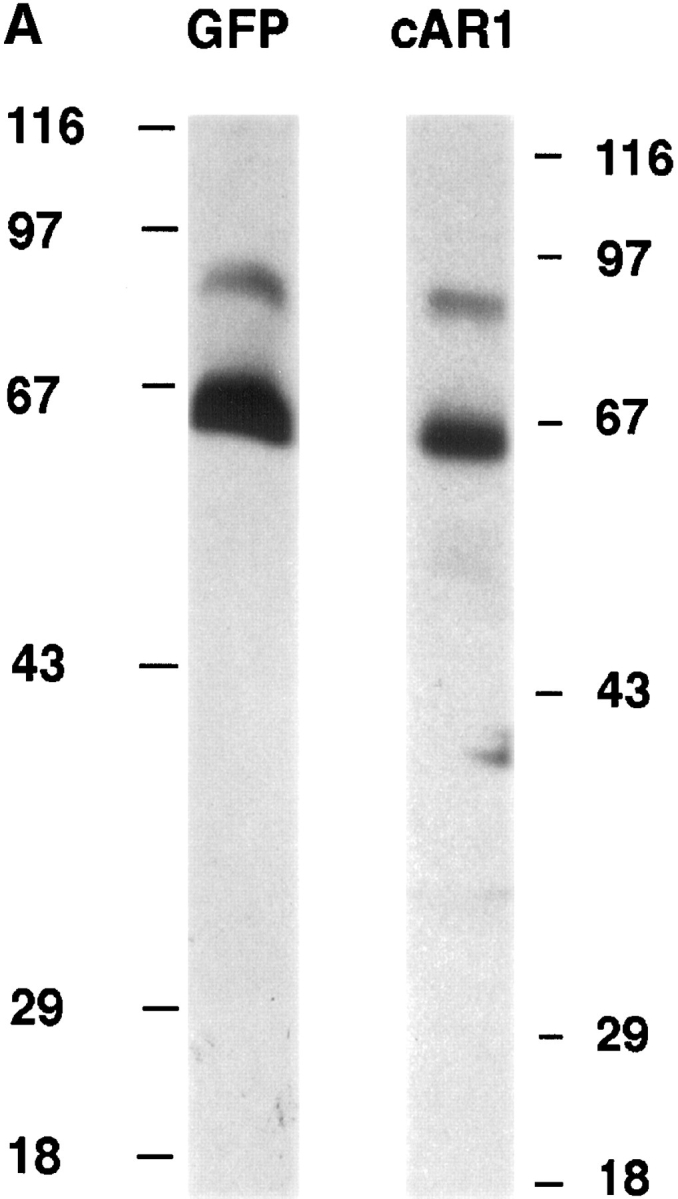
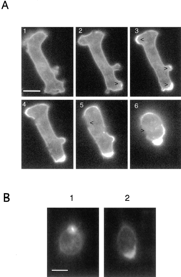
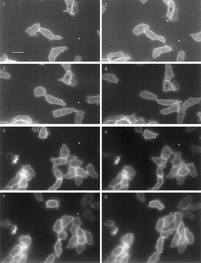
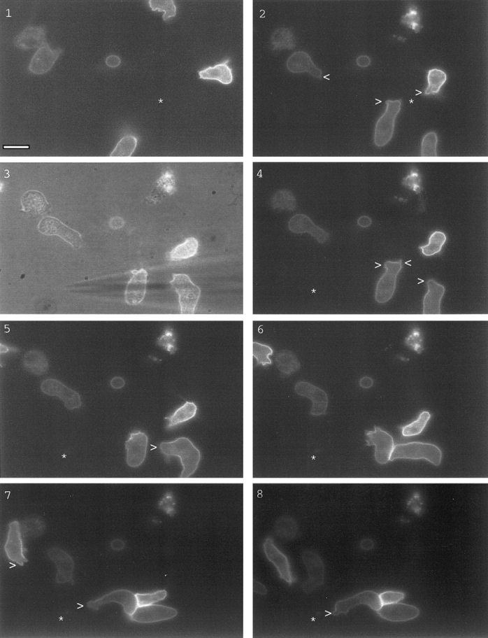
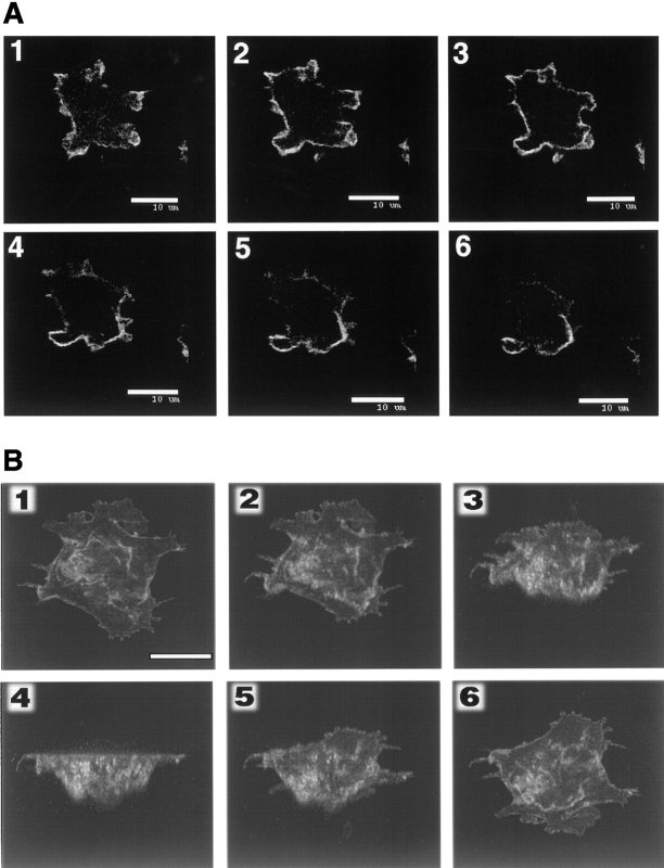
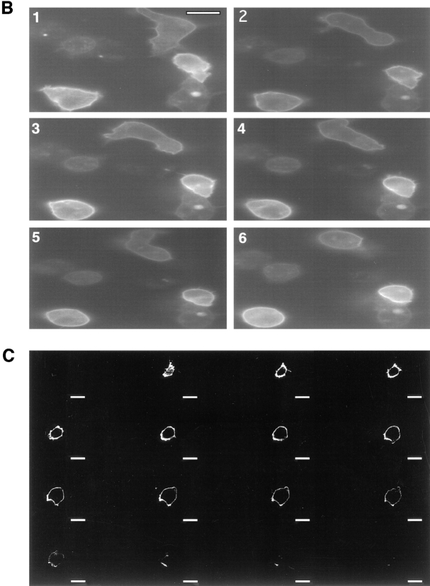
While the localization of chemoattractant receptors on randomly oriented cells has been previously studied by immunohistochemistry, the instantaneous distribution of receptors on living cells undergoing directed migration has not been determined. To do this, we replaced cAR1, the primary cAMP receptor of Dictyostelium, with a cAR1-green fluorescence protein fusion construct. We found that this chimeric protein is functionally indistinguishable from wild-type cAR1. By time-lapse imaging of single cells, we observed that the receptors remained evenly distributed on the cell surface and all of its projections during chemotaxis involving turns and reversals of polarity directed by repositioning of a chemoattractant-filled micropipet. Thus, cell polarization cannot result from a gradient-induced asymmetric distribution of chemoattractant receptors. Some newly extended pseudopods at migration fronts showed a transient drop in fluorescence signals, suggesting that the flow of receptors into these zones may slightly lag behind the protrusion process. Challenge with a uniform increase in chemoattractant, sufficient to cause a dramatic decrease in the affinity of surface binding sites and cell desensitization, also did not significantly alter the distribution profile. Hence, the induced reduction in binding activity and cellular sensitivity cannot be due to receptor relocalization. The chimeric receptors were able to "cap" rapidly during treatment with Con A, suggesting that they are mobile in the plane of the cell membrane. This capping was not influenced by pretreatment with chemoattractant.
虽然先前已通过免疫组织化学研究了趋化因子受体在随机定向细胞上的定位,但尚未确定其在经历定向迁移的活细胞上的瞬时分布。为此,我们用cAR1-绿色荧光蛋白融合构建体取代了盘基网柄菌的主要cAMP受体cAR1。我们发现这种嵌合蛋白在功能上与野生型cAR1没有区别。通过对单细胞的延时成像,我们观察到在趋化作用过程中,受体在细胞表面及其所有突起上保持均匀分布,趋化作用包括由重新定位充满趋化因子的微量移液器所引导的极性转变和逆转。因此,细胞极化不可能是由趋化因子受体的梯度诱导不对称分布导致的。迁移前沿一些新延伸的伪足显示荧光信号短暂下降,这表明受体流入这些区域的过程可能略滞后于突起过程。用均匀增加的趋化因子进行刺激,足以导致表面结合位点亲和力显著降低和细胞脱敏,也没有显著改变分布模式。因此,诱导的结合活性降低和细胞敏感性降低不可能是由于受体重新定位。在用刀豆球蛋白A处理期间,嵌合受体能够迅速“帽化”,这表明它们在细胞膜平面内是可移动的。这种帽化不受趋化因子预处理的影响。