Shimizu A, Kitamura H, Masuda Y, Ishizaki M, Sugisaki Y, Yamanaka N
Department of Pathology, Nippon Medical School, Tokyo, Japan.
Am J Pathol. 1997 Nov;151(5):1231-9.
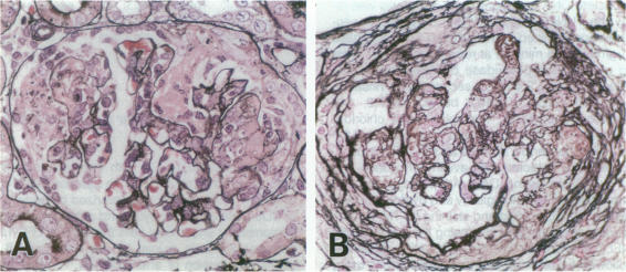
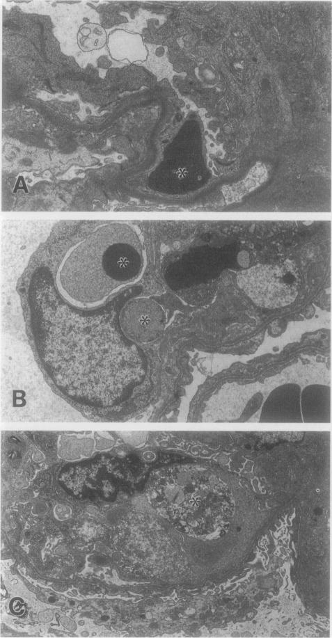
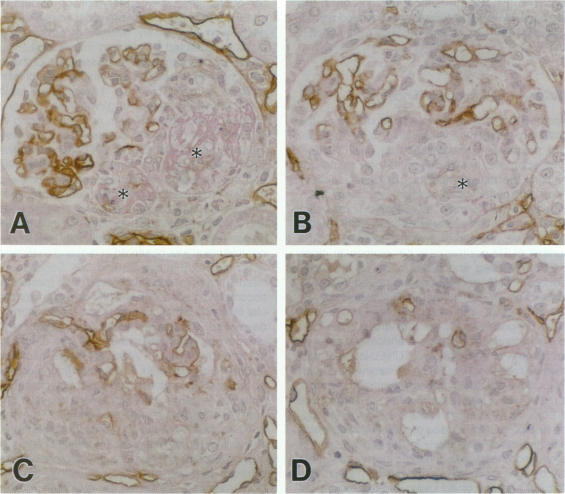
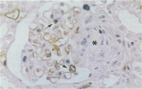
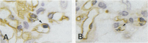
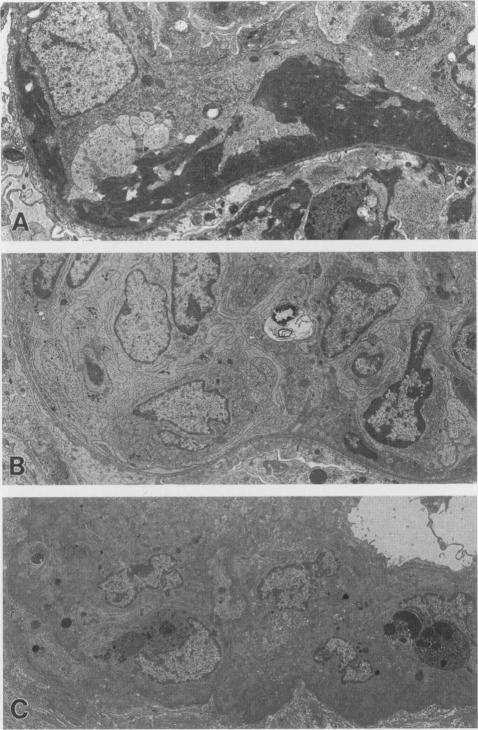
Glomerulonephritis (GN) leading to glomerular sclerosis remains an important cause of renal failure. The glomerulus is a capillary network, but endothelial and vascular reactions during progressive GN are not well understood. We have, therefore, examined the morphological alterations of glomerular capillary network and endothelial cells during the progression of damaged glomeruli to glomerular sclerosis. A progressive model of anti-glomerular basement membrane (GBM) GN was induced in Wistar-Kyoto (WKY) rats with a single injection of anti-rat GBM antibody. Severe necrotizing glomerular injuries were observed between day 5 and week 3 with a reduction in the number of total glomerular endothelial cells and total glomerular capillary lumina per glomerular cross sections. In necrotizing lesions, the glomerular endothelial cells were lost with the destruction of the glomerular capillary network. Moreover, angiogenic capillary repair with proliferation of endothelial cells was rare in severely damaged regions of glomeruli. Subsequently, mesangial hypercellularity and marked mesangial matrix accumulation occurred with absence of the development of a capillary network, and the necrotizing lesions progressed to sclerotic scars until 8 weeks. Although active necrotizing lesions could not be seen in damaged glomeruli between week 4 and week 8, the number of apoptotic endothelial cells gradually increased in the glomerular capillaries (0.10 +/- 0.01 apoptotic endothelial cells/glomerular cross section at week 8 versus 0.00 +/- 0.00 control cells (mean +/- SEM; P < 0.05) with the progression of glomerular sclerosis. Whereas the number of apoptotic endothelial cells increased in the damaged glomeruli, the number of total glomerular endothelial cells decreased (9.3 +/- 3.0 cells/glomerular cross section at week 8 versus 24.8 +/- 3.0 cells in control (mean +/- SD); P < 0.001) with regression of glomerular capillaries (3.6 +/- 2.5 capillary lumina/glomerular cross section at week 8 versus 35.0 +/- 5.0 capillary lumina in control (mean +/- SD); P < 0.001). Finally, glomerular endothelial cells could not be detected in the sclerotic lesions in progressive anti-GBM GN in WKY rats. These data indicate that the destruction of the capillary network of glomeruli and subsequent incomplete angiogenic capillary repair leads to glomerular sclerosis in progressive GN. Endothelial cell apoptosis with glomerular capillary regression may also contribute to the development of glomerular sclerosis. Injury of the glomerular capillary network with endothelial cell damage, including apoptosis and subsequent incomplete capillary repair, plays an important role in the progression of glomerular sclerosis during anti-GBM GN in WKY rats.
导致肾小球硬化的肾小球肾炎(GN)仍是肾衰竭的一个重要原因。肾小球是一个毛细血管网络,但在进行性GN过程中的内皮和血管反应尚未完全明确。因此,我们研究了受损肾小球进展为肾小球硬化过程中肾小球毛细血管网络和内皮细胞的形态学改变。通过单次注射抗大鼠肾小球基底膜(GBM)抗体,在Wistar-Kyoto(WKY)大鼠中诱导出抗GBM GN的进行性模型。在第5天至第3周期间观察到严重的坏死性肾小球损伤,每个肾小球横切面的肾小球内皮细胞总数和肾小球毛细血管腔总数减少。在坏死性病变中,随着肾小球毛细血管网络的破坏,肾小球内皮细胞丢失。此外,在肾小球严重受损区域,内皮细胞增殖的血管生成性毛细血管修复很少见。随后,出现系膜细胞增多和明显的系膜基质积聚,且无毛细血管网络形成,坏死性病变进展为硬化性瘢痕直至8周。虽然在第4周和第周8受损肾小球中未见活动性坏死性病变,但随着肾小球硬化的进展,肾小球毛细血管中凋亡内皮细胞的数量逐渐增加(第8周时为0.10±0.01个凋亡内皮细胞/肾小球横切面,而对照细胞为0.00±0.00个(平均值±SEM;P<0.05)。随着受损肾小球中凋亡内皮细胞数量增加,肾小球内皮细胞总数减少(第8周时为9.3±3.0个细胞/肾小球横切面,而对照组为24.8±3.0个细胞(平均值±SD);P<0.001),同时肾小球毛细血管退化(第8周时为3.6±2.5个毛细血管腔/肾小球横切面,而对照组为35.0±5.0个毛细血管腔(平均值±SD);P<0.001)。最终,在WKY大鼠进行性抗GBM GN的硬化性病变中未检测到肾小球内皮细胞。这些数据表明,肾小球毛细血管网络的破坏以及随后不完全的血管生成性毛细血管修复导致进行性GN中的肾小球硬化。内皮细胞凋亡伴肾小球毛细血管退化也可能促成肾小球硬化的发展。肾小球毛细血管网络损伤伴内皮细胞损伤,包括凋亡及随后不完全的毛细血管修复,在WKY大鼠抗GBM GN过程中肾小球硬化的进展中起重要作用。