Wolter K G, Hsu Y T, Smith C L, Nechushtan A, Xi X G, Youle R J
Biochemistry Section, Surgical Neurology Branch, Laboratory of Molecular Biology, National Institute of Neurological Disorders and Stroke, National Institutes of Health, Bethesda, Maryland 20892, USA.
J Cell Biol. 1997 Dec 1;139(5):1281-92. doi: 10.1083/jcb.139.5.1281.
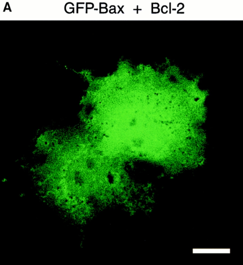
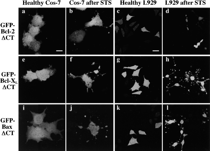
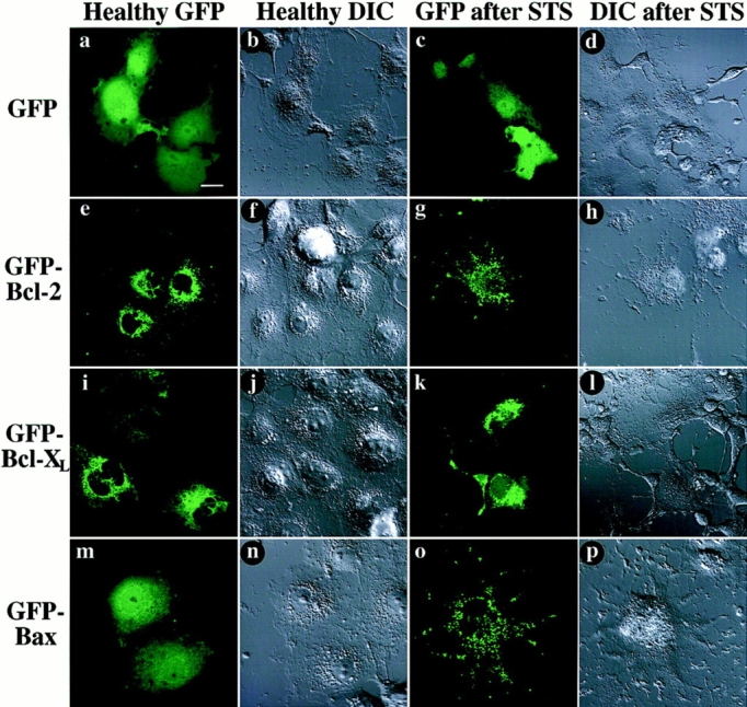
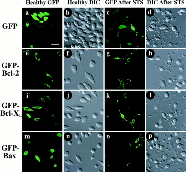
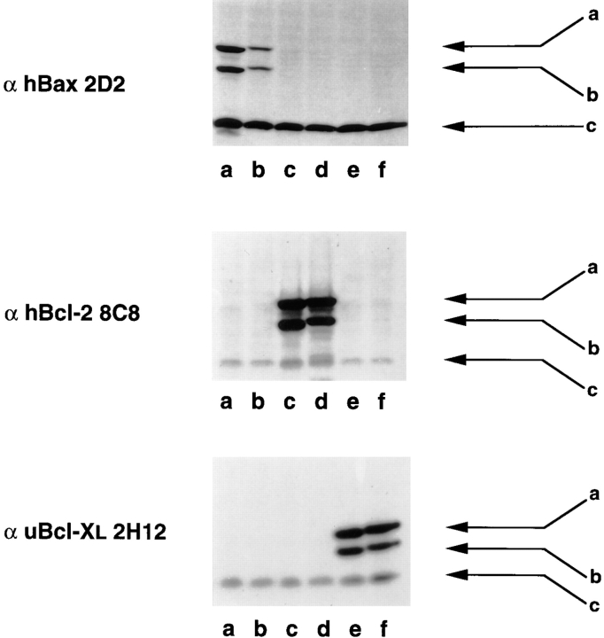
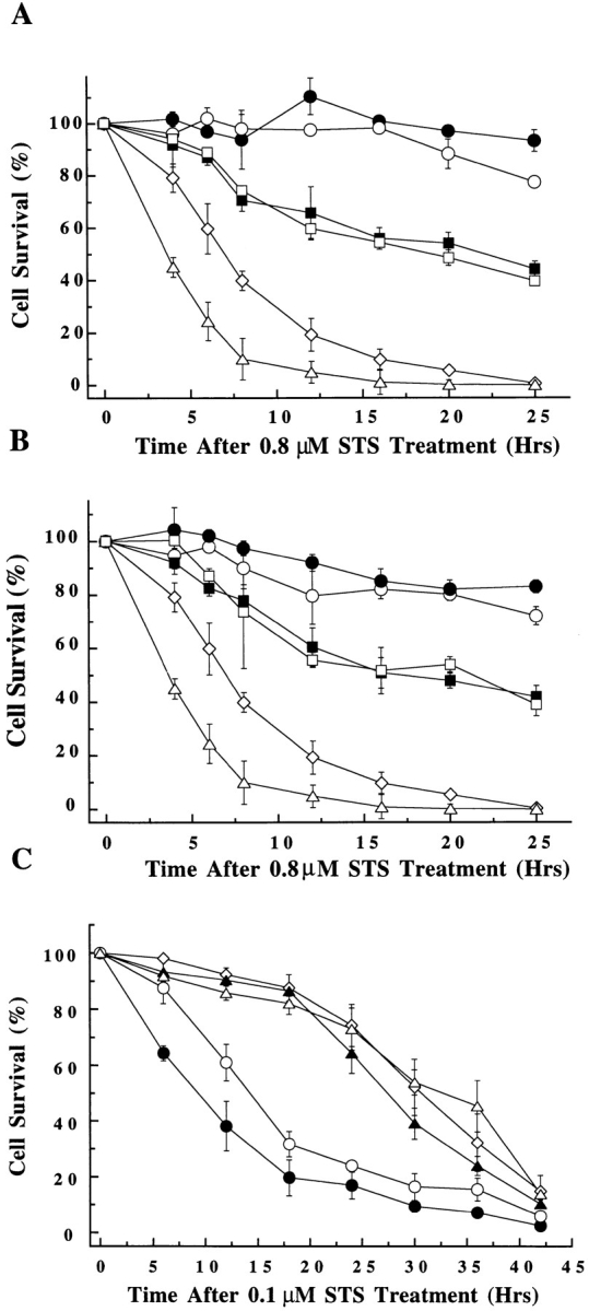
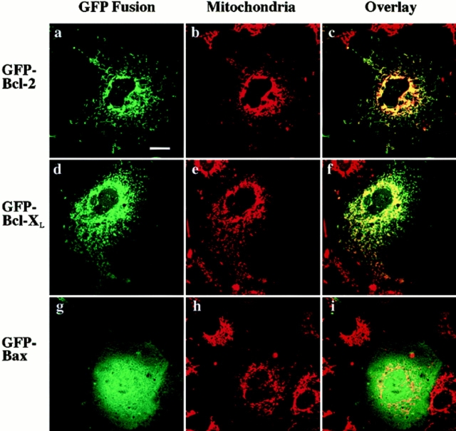
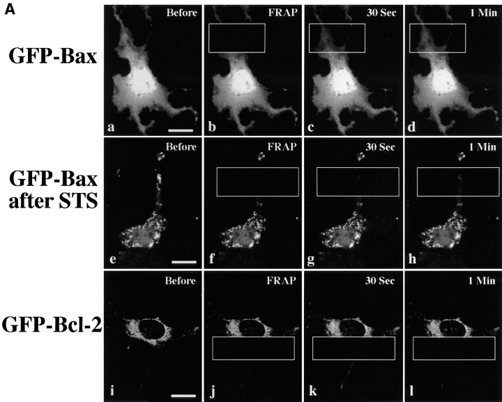
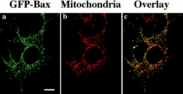
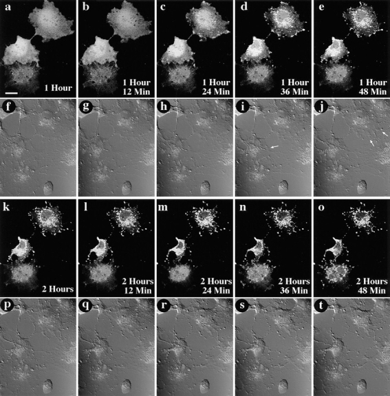
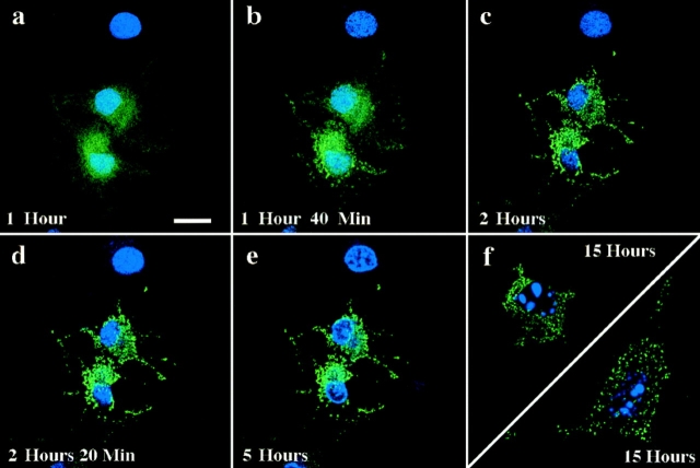
Bax, a member of the Bcl-2 protein family, accelerates apoptosis by an unknown mechanism. Bax has been recently reported to be an integral membrane protein associated with organelles or bound to organelles by Bcl-2 or a soluble protein found in the cytosol. To explore Bcl-2 family member localization in living cells, the green fluorescent protein (GFP) was fused to the NH2 termini of Bax, Bcl-2, and Bcl-XL. Confocal microscopy performed on living Cos-7 kidney epithelial cells and L929 fibroblasts revealed that GFP-Bcl-2 and GFP-Bcl-XL had a punctate distribution and colocalized with a mitochondrial marker, whereas GFP-Bax was found diffusely throughout the cytosol. Photobleaching analysis confirmed that GFP-Bax is a soluble protein, in contrast to organelle-bound GFP-Bcl-2. The diffuse localization of GFP-Bax did not change with coexpression of high levels of Bcl-2 or Bcl-XL. However, upon induction of apoptosis, GFP-Bax moved intracellularly to a punctate distribution that partially colocalized with mitochondria. Once initiated, this Bax movement was complete within 30 min, before cellular shrinkage or nuclear condensation. Removal of a COOH-terminal hydrophobic domain from GFP-Bax inhibited redistribution during apoptosis and inhibited the death-promoting activity of both Bax and GFP-Bax. These results demonstrate that in cells undergoing apoptosis, an early, dramatic change occurs in the intracellular localization of Bax, and this redistribution of soluble Bax to organelles appears important for Bax to promote cell death.
Bax是Bcl-2蛋白家族的成员之一,通过未知机制加速细胞凋亡。最近有报道称,Bax是一种整合膜蛋白,与细胞器相关联,或者通过Bcl-2与细胞器结合,或者是一种存在于胞质溶胶中的可溶性蛋白。为了探究Bcl-2家族成员在活细胞中的定位,绿色荧光蛋白(GFP)被融合到Bax、Bcl-2和Bcl-XL的NH2末端。对活的Cos-7肾上皮细胞和L929成纤维细胞进行共聚焦显微镜观察发现,GFP-Bcl-2和GFP-Bcl-XL呈点状分布,并与线粒体标记物共定位,而GFP-Bax则在整个胞质溶胶中呈弥散分布。光漂白分析证实,与结合在细胞器上的GFP-Bcl-2不同,GFP-Bax是一种可溶性蛋白。GFP-Bax的弥散定位不会因高水平的Bcl-2或Bcl-XL的共表达而改变。然而,在诱导细胞凋亡时,GFP-Bax在细胞内移动到点状分布,部分与线粒体共定位。一旦启动,这种Bax的移动在30分钟内完成,早于细胞皱缩或核浓缩。从GFP-Bax上去除COOH末端疏水结构域可抑制细胞凋亡过程中的重新分布,并抑制Bax和GFP-Bax的促死亡活性。这些结果表明,在经历细胞凋亡的细胞中,Bax的细胞内定位会发生早期的显著变化,而可溶性Bax向细胞器的这种重新分布似乎对Bax促进细胞死亡很重要。