Ellenberg J, Siggia E D, Moreira J E, Smith C L, Presley J F, Worman H J, Lippincott-Schwartz J
Cell Biology and Metabolism Branch, National Institute of Child Health and Human Development, National Institutes of Health (NIH), Bethesda, Maryland 20892, USA.
J Cell Biol. 1997 Sep 22;138(6):1193-206. doi: 10.1083/jcb.138.6.1193.
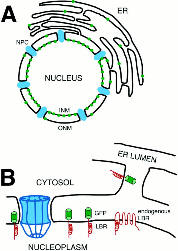
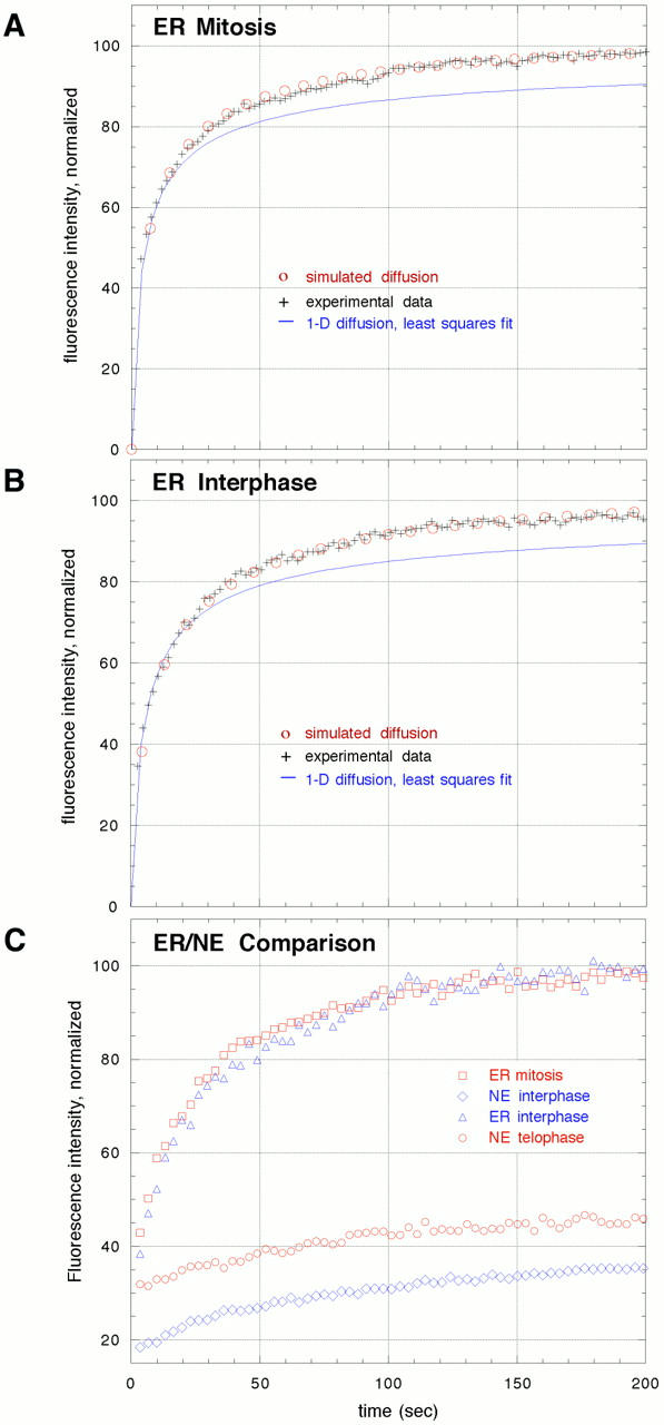
The mechanisms of localization and retention of membrane proteins in the inner nuclear membrane and the fate of this membrane system during mitosis were studied in living cells using the inner nuclear membrane protein, lamin B receptor, fused to green fluorescent protein (LBR-GFP). Photobleaching techniques revealed the majority of LBR-GFP to be completely immobilized in the nuclear envelope (NE) of interphase cells, suggesting a tight binding to heterochromatin and/or lamins. A subpopulation of LBR-GFP within ER membranes, by contrast, was entirely mobile and diffused rapidly and freely (D = 0. 41 +/- 0.1 microm2/s). High resolution confocal time-lapse imaging in mitotic cells revealed LBR-GFP redistributing into the interconnected ER membrane system in prometaphase, exhibiting the same high mobility and diffusion constant as observed in interphase ER membranes. LBR-GFP rapidly diffused across the cell within the membrane network defined by the ER, suggesting the integrity of the ER was maintained in mitosis, with little or no fragmentation and vesiculation. At the end of mitosis, nuclear membrane reformation coincided with immobilization of LBR-GFP in ER elements at contact sites with chromatin. LBR-GFP-containing ER membranes then wrapped around chromatin over the course of 2-3 min, quickly and efficiently compartmentalizing nuclear material. Expansion of the NE followed over the course of 30-80 min. Thus, selective changes in lateral mobility of LBR-GFP within the ER/NE membrane system form the basis for its localization to the inner nuclear membrane during interphase. Such changes, rather than vesiculation mechanisms, also underlie the redistribution of this molecule during NE disassembly and reformation in mitosis.
利用与绿色荧光蛋白融合的内核膜蛋白——核纤层蛋白B受体(LBR-GFP),在活细胞中研究了膜蛋白在内核膜中的定位和滞留机制,以及该膜系统在有丝分裂期间的命运。光漂白技术显示,大多数LBR-GFP完全固定在间期细胞的核膜(NE)中,这表明它与异染色质和/或核纤层蛋白紧密结合。相比之下,内质网(ER)膜内的一小部分LBR-GFP完全可移动,且快速自由扩散(扩散系数D = 0.41±0.1μm2/s)。对有丝分裂细胞进行的高分辨率共聚焦延时成像显示,LBR-GFP在前中期重新分布到相互连接的内质网系统中,其迁移率和扩散常数与间期内质网中观察到的相同。LBR-GFP在由内质网界定的膜网络内迅速扩散穿过细胞,这表明内质网在有丝分裂期间保持完整,几乎没有或没有碎片化和囊泡化。有丝分裂末期,核膜重新形成与LBR-GFP在内质网元件与染色质接触部位的固定同时发生。然后,含有LBR-GFP的内质网膜在2-3分钟内包裹染色质,迅速有效地分隔核物质。随后,核膜在30-80分钟内扩展。因此,LBR-GFP在内质网/核膜系统内横向迁移率的选择性变化是其在间期定位于内核膜的基础。这些变化而非囊泡化机制,也是该分子在有丝分裂期间核膜解体和重新形成过程中重新分布的基础。