Sarks S H
Br J Ophthalmol. 1976 May;60(5):324-41. doi: 10.1136/bjo.60.5.324.
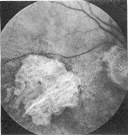
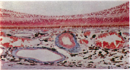
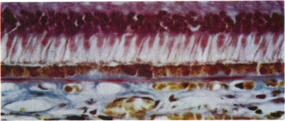
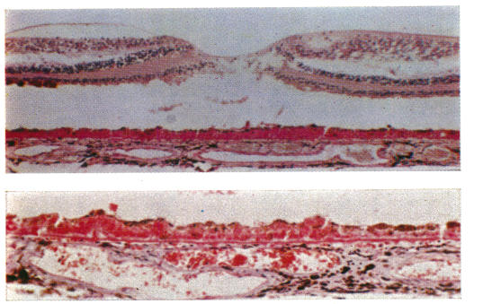
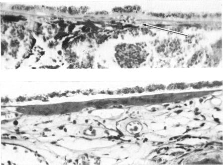
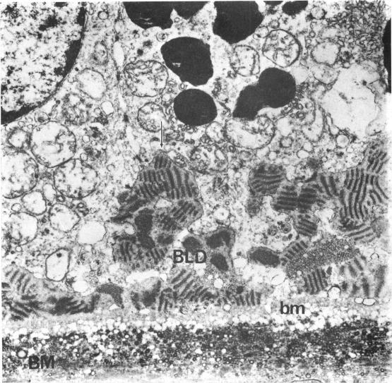
Clinical and pathological examination was performed on 378 eyes from 216 patients aged 43 to 97 years. This series represented eyes in which the fundi were normal or showed various manifestations of senile macular degeneration. The eyes were divided into six groups according to the histological appearance of a linear deposit at the base of the retinal pigment cells. Groups I and II were considered to represent normal ageing, Groups III and IV the progressive development of senile macular degeneration and Groups V and VI the end-results. Group I showed no basal linear deposit. Thickening and hyalinization of Bruch's membrane was noted as early as the fifth decade. Group II showed patchy development of the basal linear deposit in relation to thickened or basophilic segments of Bruch's membrane, or over intercapillary hyalinization extending to the level of the outer surface of the choriocapillaris. Almost all eyes in these two groups retained a normal fundus appearance but visual acuity declined with age even in the absence of other causes. In Group III the basal deposit formed a thin continuous layer associated with moderate degeneration of the retinal pigment epithelium. More than half the eyes had developed a clinical disturbance of pigmentation and in most vision was reduced. Group IV was characterized by thickening of the deposit and more pronounced disturbance of the pigment epithelium. Clinically most eyes showed coarse pigmentary changes and vision was in the order of 6/24. 14-3 per cent of eyes in this group showed early neovascularization from the choroid. In Group V the pigment epithelium disappeared to produce circumscribed areas of depigmentation. The basal linear deposit could be traced throughout the depigmented area in most eyes. Thin fibrovascular sheets were found beneath the pigment epithelium in 41-7 per cent of eyes. Group VI represented disciform degeneration. The basal linear deposit could often be demonstrated as a disrupted hyalinized layer incorporated into the scar. Disciform degeneration was an alternative end-result to geographical atrophy. In each group the clinical and histological findings may be modified by the presence of drusen or by atrophy of the choroid. The basal linear deposit consisted of banded fibres embedded in granular material lying between the plasma infoldings and the basement membrane of the retinal pigment epithelium. This deposit seems to be a manifestation of gradual failure of the pigment epithelium and proved to be the most suitable criterion by which to study the natural history of senile macular degeneration.
对216例年龄在43至97岁患者的378只眼睛进行了临床和病理检查。该系列代表眼底正常或表现出各种老年性黄斑变性表现的眼睛。根据视网膜色素细胞底部线性沉积物的组织学外观,将眼睛分为六组。第一组和第二组被认为代表正常衰老,第三组和第四组代表老年性黄斑变性的进展,第五组和第六组代表最终结果。第一组未显示基底线性沉积物。早在第五个十年就注意到了布鲁赫膜的增厚和玻璃样变性。第二组显示基底线性沉积物呈斑片状发展,与布鲁赫膜增厚或嗜碱性节段有关,或在延伸至脉络膜毛细血管外表面水平的毛细血管间玻璃样变性之上。这两组中的几乎所有眼睛眼底外观均保持正常,但即使在没有其他原因的情况下,视力也会随着年龄的增长而下降。在第三组中,基底沉积物形成一层薄的连续层,与视网膜色素上皮的中度变性有关。超过一半的眼睛出现了色素沉着的临床紊乱,大多数眼睛的视力下降。第四组的特征是沉积物增厚,色素上皮的紊乱更明显。临床上,大多数眼睛表现出粗大的色素变化,视力约为6/24。该组中14.3%的眼睛显示出脉络膜早期新生血管形成。在第五组中,色素上皮消失,形成局限性色素脱失区域。在大多数眼睛中,基底线性沉积物可在整个色素脱失区域追踪到。41.7%的眼睛在色素上皮下方发现了薄的纤维血管片层。第六组代表盘状变性。基底线性沉积物通常可显示为融入瘢痕中的破碎玻璃样变性层。盘状变性是地图状萎缩的另一种最终结果。在每组中,临床和组织学发现可能会因玻璃膜疣的存在或脉络膜萎缩而改变。基底线性沉积物由嵌入视网膜色素上皮细胞浆褶皱和基底膜之间颗粒物质中的带状纤维组成。这种沉积物似乎是色素上皮逐渐衰竭的表现,并且被证明是研究老年性黄斑变性自然史的最合适标准。