Jeffers M D, Milton J, Herriot R, McKean M
Department of Cytopathology, Aberdeen Royal Infirmary, UK.
J Clin Pathol. 1998 Mar;51(3):189-96. doi: 10.1136/jcp.51.3.189.
To assess the value of flow cytometry (FCM) in the diagnosis and classification of reactive lymphoid hyperplasia and malignant lymphoma by fine needle aspiration (FNA) cytology.
Forty six fine needle aspirates of lymphoproliferative disorders were examined by FCM as well as routine cytological assessment. An immunoglobulin light chain ratio (LCR) was calculated for clonality analysis. Additional immunophenotyping was performed in 15 cases.
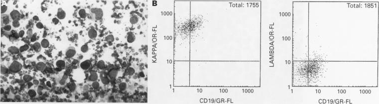
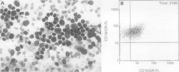
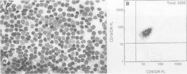
All 25 cases of reactive lymphoid hyperplasia were polyclonal by FCM (LCR < 2/1); 17 of 20 cases of B cell non-Hodgkin's lymphoma were monoclonal (LCR > 3/1). Analysis of cells based on size facilitated detection of small populations of clonal neoplastic cells. Analysis of CD5, CD10, and CD23 expression by FCM facilitated subclassification of mantle cell lymphoma, small lymphocytic lymphoma, and some lymphomas of follicle centre cell origin. One case of T cell non-Hodgkin's lymphoma was correctly classified by FCM.
FNA cytology is a reliable method for investigation of lymphoproliferative disorders. Although excision biopsy and histopathological examination remain the gold standard for primary diagnosis and classification of non-Hodgkin's lymphoma, FNA cytology with clonality analysis and immunophenotyping by FCM is useful for distinguishing reactive from neoplastic lymphoid populations, and can facilitate lymphoma classification.
通过细针穿刺(FNA)细胞学评估流式细胞术(FCM)在反应性淋巴组织增生和恶性淋巴瘤诊断及分类中的价值。
对46例淋巴增生性疾病的细针穿刺样本进行FCM检测以及常规细胞学评估。计算免疫球蛋白轻链比值(LCR)用于克隆性分析。另外对15例进行了免疫表型分析。
FCM检测显示,所有25例反应性淋巴组织增生均为多克隆性(LCR<2/1);20例B细胞非霍奇金淋巴瘤中有17例为单克隆性(LCR>3/1)。基于细胞大小的分析有助于检测少量克隆性肿瘤细胞。通过FCM分析CD5、CD10和CD23表达有助于套细胞淋巴瘤、小淋巴细胞淋巴瘤及部分滤泡中心细胞源性淋巴瘤的亚分类。1例T细胞非霍奇金淋巴瘤通过FCM正确分类。
FNA细胞学是研究淋巴增生性疾病的可靠方法。虽然切除活检和组织病理学检查仍是非霍奇金淋巴瘤初步诊断和分类的金标准,但结合克隆性分析和FCM免疫表型分析的FNA细胞学有助于区分反应性和肿瘤性淋巴细胞群,并可促进淋巴瘤的分类。