Senzel L, Huynh P D, Jakes K S, Collier R J, Finkelstein A
Department of Neuroscience, Albert Einstein College of Medicine, Bronx, New York 10461, USA.
J Gen Physiol. 1998 Sep;112(3):317-24. doi: 10.1085/jgp.112.3.317.
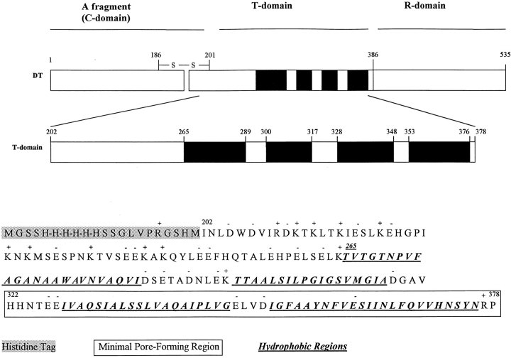
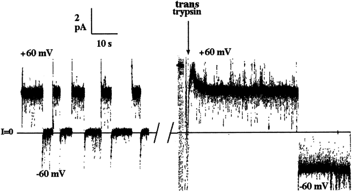
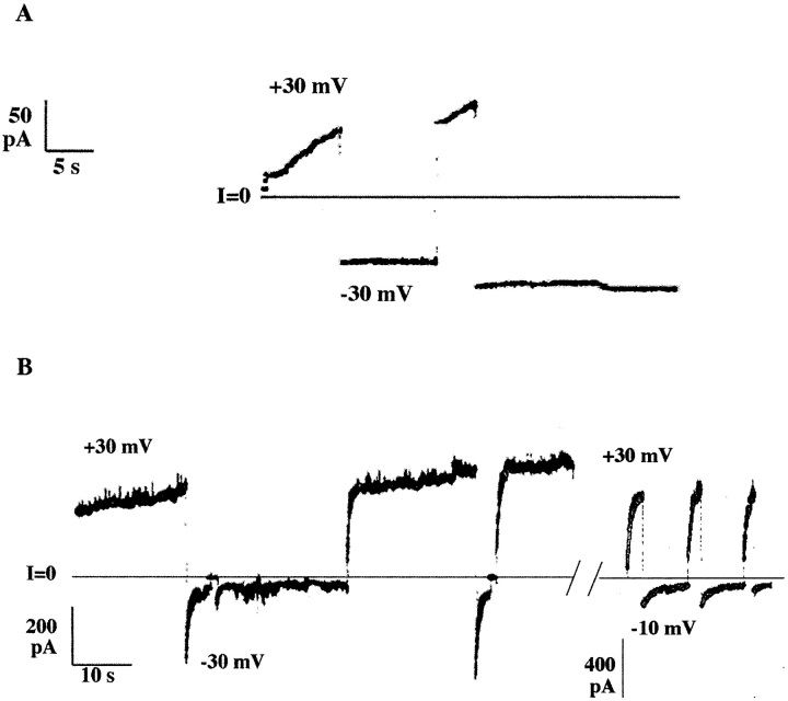
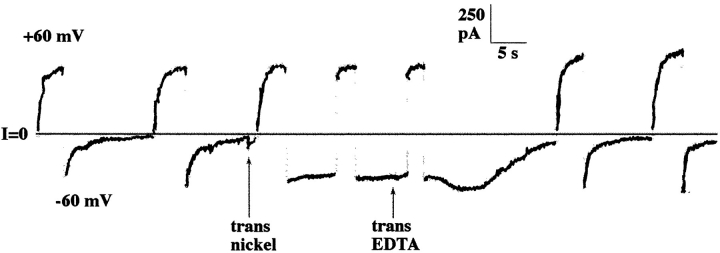
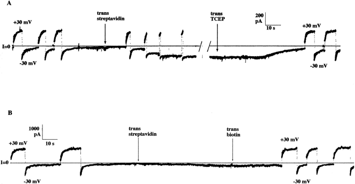
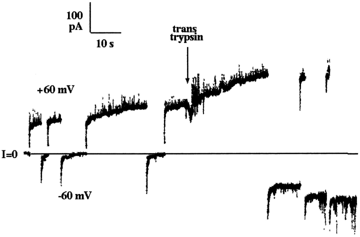
The T domain of diphtheria toxin, which extends from residue 202 to 378, causes the translocation of the catalytic A fragment (residues 1-201) across endosomal membranes and also forms ion-conducting channels in planar phospholipid bilayers. The carboxy terminal 57-amino acid segment (322-378) in the T domain is all that is required to form these channels, but its ability to do so is greatly augmented by the portion of the T domain upstream from this. In this work, we show that in association with channel formation by the T domain, its NH2 terminus, as well as some or all of the adjacent hydrophilic 63 amino acid segment, cross the lipid bilayer. The phenomenon that enabled us to demonstrate that the NH2-terminal region of the T domain was translocated across the membrane was the rapid closure of channels at cis negative voltages when the T domain contained a histidine tag at its NH2 terminus. The inhibition of this effect by trans nickel, and by trans streptavidin when the histidine tag sequence was biotinylated, clearly established that the histidine tag was present on the trans side of the membrane. Furthermore, the inhibition of rapid channel closure by trans trypsin, combined with mutagenesis to localize the trypsin site, indicated that some portion of the 63 amino acid NH2-terminal segment of the T domain was also translocated to the trans side of the membrane. If the NH2 terminus was forced to remain on the cis side, by streptavidin binding to the biotinylated histidine tag sequence, channel formation was severely disrupted. Thus, normal channel formation by the T domain requires that its NH2 terminus be translocated across the membrane from the cis to the trans side, even though the NH2 terminus is >100 residues removed from the channel-forming part of the molecule.
白喉毒素的T结构域(从第202位氨基酸延伸至378位氨基酸)可促使催化性A片段(第1 - 201位氨基酸)穿过内体膜,并且还能在平面磷脂双分子层中形成离子传导通道。T结构域的羧基末端57个氨基酸片段(322 - 378)是形成这些通道所必需的全部,但T结构域中该片段上游部分极大地增强了其形成通道的能力。在这项研究中,我们表明,与T结构域形成通道相关联的是,其氨基末端以及部分或全部相邻的63个亲水性氨基酸片段会穿过脂质双分子层。当T结构域在其氨基末端含有组氨酸标签时,顺式负电压下通道的快速关闭这一现象使我们能够证明T结构域的氨基末端区域穿过了膜。反式镍以及当组氨酸标签序列被生物素化时反式链霉亲和素对这种效应的抑制,明确证实了组氨酸标签位于膜的反式侧。此外,反式胰蛋白酶对快速通道关闭的抑制作用,结合诱变来定位胰蛋白酶作用位点,表明T结构域的63个氨基酸氨基末端片段的某些部分也转移到了膜的反式侧。如果通过链霉亲和素与生物素化的组氨酸标签序列结合迫使氨基末端留在顺式侧,通道形成会受到严重破坏。因此,T结构域正常的通道形成要求其氨基末端从顺式侧穿过膜转移到反式侧,尽管氨基末端距离分子的通道形成部分超过100个残基。