• 文献检索
  • 文档翻译
  • 深度研究
  • 学术资讯
  • Suppr Zotero 插件Zotero 插件
  • 邀请有礼
  • 套餐&价格
  • 历史记录
应用&插件
Suppr Zotero 插件Zotero 插件浏览器插件Mac 客户端Windows 客户端微信小程序
定价
高级版会员购买积分包购买API积分包
服务
文献检索文档翻译深度研究API 文档MCP 服务
关于我们
关于 Suppr公司介绍联系我们用户协议隐私条款
关注我们

Suppr 超能文献

核心技术专利:CN118964589B侵权必究
粤ICP备2023148730 号-1Suppr @ 2026

文献检索

告别复杂PubMed语法,用中文像聊天一样搜索,搜遍4000万医学文献。AI智能推荐,让科研检索更轻松。

立即免费搜索

文件翻译

保留排版,准确专业,支持PDF/Word/PPT等文件格式,支持 12+语言互译。

免费翻译文档

深度研究

AI帮你快速写综述,25分钟生成高质量综述,智能提取关键信息,辅助科研写作。

立即免费体验

人类食管中的朗格汉斯细胞。

Langerhans cells in the human oesophagus.

作者信息

Yassin T M, Toner P G

出版信息

J Anat. 1976 Nov;122(Pt 2):435-45.

PMID:1002612
原文链接:https://pmc.ncbi.nlm.nih.gov/articles/PMC1231913/
Abstract

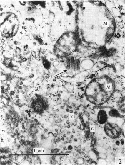
The dendrite cells of Langerhans, first identified in the epidermis, have now been observed in the middle and superficial layers of the normal human oesophageal mucosa. They exhibit typical Langerhans granules, but no desmosomes and tonofilaments. They often have irregular indented nuclei, with a relatively pale cytoplasm contrasting with that of the adjacent squamous cells. These cells are sometimes difficult to distinguish from intra-epithelial lymphocytes, which are also encountered in the oesophageal mucosa and which share certain ultrastructural characteristics with Langerhans cells.

摘要

朗格汉斯树突状细胞最初在表皮中被识别,现在已在正常人类食管黏膜的中层和表层被观察到。它们呈现出典型的朗格汉斯颗粒,但没有桥粒和张力丝。它们的细胞核常呈不规则凹陷状,与相邻鳞状细胞相比,细胞质相对较淡。这些细胞有时难以与上皮内淋巴细胞区分开来,上皮内淋巴细胞在食管黏膜中也会出现,并且与朗格汉斯细胞具有某些超微结构特征。

https://cdn.ncbi.nlm.nih.gov/pmc/blobs/7296/1231913/1cf96036fbee/janat00369-0225-a.jpg
https://cdn.ncbi.nlm.nih.gov/pmc/blobs/7296/1231913/c9f7b3b9d9f0/janat00369-0219-a.jpg
https://cdn.ncbi.nlm.nih.gov/pmc/blobs/7296/1231913/2f8ada55fb78/janat00369-0221-a.jpg
https://cdn.ncbi.nlm.nih.gov/pmc/blobs/7296/1231913/473039f7d61b/janat00369-0221-b.jpg
https://cdn.ncbi.nlm.nih.gov/pmc/blobs/7296/1231913/a11fbff419c2/janat00369-0222-a.jpg
https://cdn.ncbi.nlm.nih.gov/pmc/blobs/7296/1231913/e0bd0ea331cc/janat00369-0222-b.jpg
https://cdn.ncbi.nlm.nih.gov/pmc/blobs/7296/1231913/53c73e568cef/janat00369-0223-a.jpg
https://cdn.ncbi.nlm.nih.gov/pmc/blobs/7296/1231913/3d0a8aad1dff/janat00369-0223-b.jpg
https://cdn.ncbi.nlm.nih.gov/pmc/blobs/7296/1231913/6e15994ce8d7/janat00369-0224-a.jpg
https://cdn.ncbi.nlm.nih.gov/pmc/blobs/7296/1231913/b0bd780df847/janat00369-0224-b.jpg
https://cdn.ncbi.nlm.nih.gov/pmc/blobs/7296/1231913/1cf96036fbee/janat00369-0225-a.jpg

相似文献

1
Langerhans cells in the human oesophagus.人类食管中的朗格汉斯细胞。
J Anat. 1976 Nov;122(Pt 2):435-45.
2
Langerhans cells in the human oviduct mucosa.人输卵管黏膜中的朗格汉斯细胞。
Ital J Anat Embryol. 1998;103(4 Suppl 1):253-8.
3
[Melanocytes, Langerhans and Merkel cells in oral epithelium].[口腔上皮中的黑素细胞、朗格汉斯细胞和默克尔细胞]
Acta Otolaryngol. 1975 Sep-Oct;80(3-4):301-11.
4
Ultrastructural observations of the oral mucosa in Anderson-Fabry disease.安德森-法布里病口腔黏膜的超微结构观察
J Oral Pathol. 1975 Nov;4(5):272-90.
5
Cells in peripheral leg lymph of normal men.正常男性腿部外周淋巴中的细胞。
Lymphology. 1978 Dec;11(4):202-7.
6
Fine structure of squamous epitheilum and submucosal glands of human oesophagus.人食管鳞状上皮和黏膜下腺的精细结构
J Anat. 1977 Jul;123(Pt 3):705-21.
7
Specialized metaplastic columnar epithelium in Barrett's esophagus. A comparative transmission electron microscopic study.巴雷特食管中的特殊化生柱状上皮。一项比较性透射电子显微镜研究。
Lab Invest. 1989 Mar;60(3):418-32.
8
Ultrastructural identification of Langerhans cells in normal swine epidermis.正常猪表皮中朗格汉斯细胞的超微结构鉴定
J Anat. 1991 Dec;179:43-6.
9
Langerhans' cells in urticaria: an electron microscopic study.荨麻疹中的朗格汉斯细胞:一项电子显微镜研究。
Indian J Dermatol. 1989 Sep;34(3):54-8.
10
Ultrastructural determination of gingival Langerhans cells in alloxan-induced diabetic rats.四氧嘧啶诱导的糖尿病大鼠牙龈朗格汉斯细胞的超微结构测定
Cell Biochem Funct. 2005 May-Jun;23(3):181-7. doi: 10.1002/cbf.1138.

引用本文的文献

1
Eosinophilic esophagitis in adults: an emerging disease.成人嗜酸性粒细胞性食管炎:一种新出现的疾病。
Dig Dis Sci. 2004 Nov-Dec;49(11-12):1884-8. doi: 10.1007/s10620-004-9588-x.
2
Langerhans cell-like dendritic cells in the cornea, tongue and oesophagus of the chicken (Gallus gallus).鸡(原鸡)角膜、舌和食管中的朗格汉斯细胞样树突状细胞。
Histochem J. 2002 Oct;34(10):507-15. doi: 10.1023/a:1024714107373.
3
Lymphocytes and Langerhans cells in the human oesophageal epithelium.人食管上皮中的淋巴细胞和朗格汉斯细胞。

本文引用的文献

1
My conception of cellular nevi.我对细胞痣的概念。
Cancer. 1951 Jan;4(1):9-38. doi: 10.1002/1097-0142(195101)4:1<9::aid-cncr2820040104>3.0.co;2-0.
2
THE LANGERHANS CELL.朗格汉斯细胞。
J Invest Dermatol. 1965 Mar;44:201-12. doi: 10.1038/jid.1965.35.
3
OBSERVATIONS ON CYTOPLASMIC ORGANELLES IN LANGERHANS CELLS OF HUMAN EPIDERMIS.人表皮朗格汉斯细胞中细胞质细胞器的观察
Virchows Arch A Pathol Anat Histopathol. 1983;401(1):45-55. doi: 10.1007/BF00644788.
4
Antigen presenting cells in situ: their identification and involvement in immunopathology.原位抗原呈递细胞:它们的鉴定及其在免疫病理学中的作用。
Clin Exp Immunol. 1983 Sep;53(3):513-20.
5
Immunological and histochemical analysis of regional variations of epidermal Langerhans cells in normal human skin.正常人皮肤表皮朗格汉斯细胞区域差异的免疫和组织化学分析。
Histochem J. 1984 May;16(5):507-19. doi: 10.1007/BF01041351.
6
S-100 protein-positive Langerhans cells in various human lung cancers, especially in peripheral adenocarcinomas.在各种人类肺癌中,尤其是外周腺癌中存在S-100蛋白阳性的朗格汉斯细胞。
Virchows Arch A Pathol Anat Histopathol. 1985;407(2):177-89. doi: 10.1007/BF00737075.
7
Does neoplasia in-situ develop due to the interaction of Epstein-Barr virus or herpes simplex virus-2 with Langerhans cells in the epithelium?原位肿瘤的发生是由于爱泼斯坦-巴尔病毒或单纯疱疹病毒-2与上皮中的朗格汉斯细胞相互作用所致吗?
Virus Genes. 1988 Jul;1(4):385-97. doi: 10.1007/BF00257101.
8
Experimental chlamydial keratitis in rabbits. Correlation with chlamydia infected McCoy tissue culture cells.兔实验性衣原体角膜炎。与衣原体感染的 McCoy 组织培养细胞的相关性。
Doc Ophthalmol. 1988 Aug;69(4):353-69. doi: 10.1007/BF00162749.
9
Human oesophageal submucosal glands. Their detection mucin, enzyme and secretory protein content.人食管黏膜下腺。其检测黏蛋白、酶和分泌蛋白含量。
Histochemistry. 1986;86(1):107-12. doi: 10.1007/BF00492353.
10
Adenosine triphosphatase-positive Langerhans-like cells in the epidermis of the chicken (Gallus gallus).鸡(原鸡)表皮中三磷酸腺苷酶阳性的朗格汉斯样细胞。
J Anat. 1991 Jun;176:1-8.
J Anat. 1964 Apr;98(Pt 2):265-70.
4
Langerhans' cell granules in histiocytosis X. The epidermal Langerhans' cell as a macrophage.组织细胞增多症X中的朗格汉斯细胞颗粒。作为巨噬细胞的表皮朗格汉斯细胞。
Arch Dermatol. 1967 Sep;96(3):298-304.
5
Skin ultrastructure in Hand-Schüller-Christian disease. Report on abnormal Langerhans' cells.汉-许-克病的皮肤超微结构。关于异常朗格汉斯细胞的报告。
Arch Dermatol. 1969 Sep;100(3):342-9.
6
Langerhans cells in a localized lesion of the eosinophilic granuloma type.嗜酸性肉芽肿型局部病变中的朗格汉斯细胞。
Lab Invest. 1969 May;20(5):412-23.
7
Electron microscope observations on Langerhans cells in the cervix.子宫颈朗格汉斯细胞的电子显微镜观察
Am J Obstet Gynecol. 1968 Oct 1;102(3):397-403. doi: 10.1016/0002-9378(68)90012-4.
8
Lymph node ultrastructure in Hand-Schüller-Christian disease.汉-许-克病中的淋巴结超微结构
Cancer. 1971 Apr;27(4):956-64. doi: 10.1002/1097-0142(197104)27:4<956::aid-cncr2820270431>3.0.co;2-g.
9
Langerhans cell granules in human hyperplastic lymph nodes.人类增生性淋巴结中的朗格汉斯细胞颗粒。
Arch Pathol. 1971 Jul;92(1):46-52.
10
Langerhans cells: uptake of tritiated thymidine.朗格汉斯细胞:氚标记胸腺嘧啶核苷的摄取
Science. 1967 Jul 28;157(3787):439-40. doi: 10.1126/science.157.3787.439.