Telzer B R, Moses M J, Rosenbaum J L
Proc Natl Acad Sci U S A. 1975 Oct;72(10):4023-7. doi: 10.1073/pnas.72.10.4023.
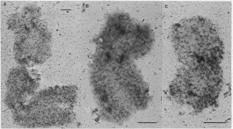
The kinetochores of isolated HeLa cell chromosomes attached to an electron microscope specimen grid, fixed in formaldehyde, and stained with alcoholic phosphotungstic acid are visible as dark, preferentially stained structures distinct from the chromatin with which they are associated. When unfixed chromosomes are immobilized by attachment to grids and incubated with chick brain tubulin, microtubules are observed to assemble onto the kinetochores. This demonstrates the competence of kinetochores in isolated chromosomes to act in vitro as microtubule assembly sites and suggests that they also possess this capacity in vivo. In addition, the results provide a possible means for isolating and characterizing kinetochores.
将分离出的HeLa细胞染色体的动粒附着在电子显微镜标本网上,用甲醛固定,并用乙醇磷钨酸染色,可见其为深色的、优先染色的结构,与它们所关联的染色质不同。当未固定的染色体通过附着在网上而固定,并与鸡脑微管蛋白一起孵育时,可观察到微管在动粒上组装。这证明了分离染色体中的动粒在体外作为微管组装位点的能力,并表明它们在体内也具有这种能力。此外,这些结果为分离和鉴定动粒提供了一种可能的方法。