• 文献检索
  • 文档翻译
  • 深度研究
  • 学术资讯
  • Suppr Zotero 插件Zotero 插件
  • 邀请有礼
  • 套餐&价格
  • 历史记录
应用&插件
Suppr Zotero 插件Zotero 插件浏览器插件Mac 客户端Windows 客户端微信小程序
定价
高级版会员购买积分包购买API积分包
服务
文献检索文档翻译深度研究API 文档MCP 服务
关于我们
关于 Suppr公司介绍联系我们用户协议隐私条款
关注我们

Suppr 超能文献

核心技术专利:CN118964589B侵权必究
粤ICP备2023148730 号-1Suppr @ 2026

文献检索

告别复杂PubMed语法,用中文像聊天一样搜索,搜遍4000万医学文献。AI智能推荐,让科研检索更轻松。

立即免费搜索

文件翻译

保留排版,准确专业,支持PDF/Word/PPT等文件格式,支持 12+语言互译。

免费翻译文档

深度研究

AI帮你快速写综述,25分钟生成高质量综述,智能提取关键信息,辅助科研写作。

立即免费体验

有丝分裂机制研究简史

A Brief History of Research on Mitotic Mechanisms.

作者信息

McIntosh J Richard, Hays Thomas

机构信息

Department of Molecular, Cellular and Developmental Biology, University of Colorado, Boulder, CO 80309, USA.

Department of Genetics, Cell Biology and Development, Medical School and College of Biological Sciences, University of Minnesota, Saint Paul, MN 55455, USA.

出版信息

Biology (Basel). 2016 Dec 21;5(4):55. doi: 10.3390/biology5040055.

DOI:10.3390/biology5040055
PMID:28009830
原文链接:https://pmc.ncbi.nlm.nih.gov/articles/PMC5192435/
Abstract

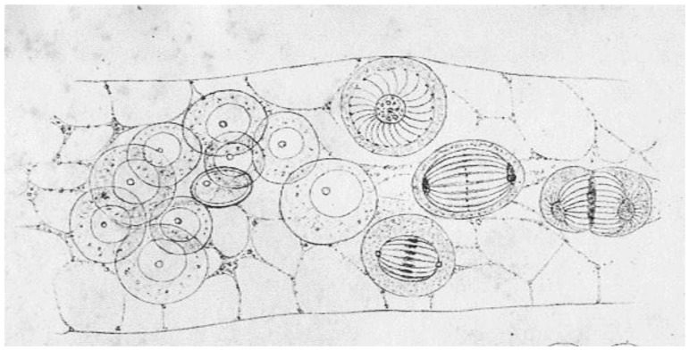
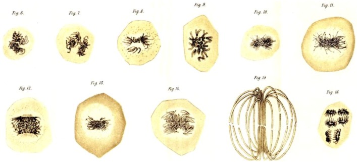
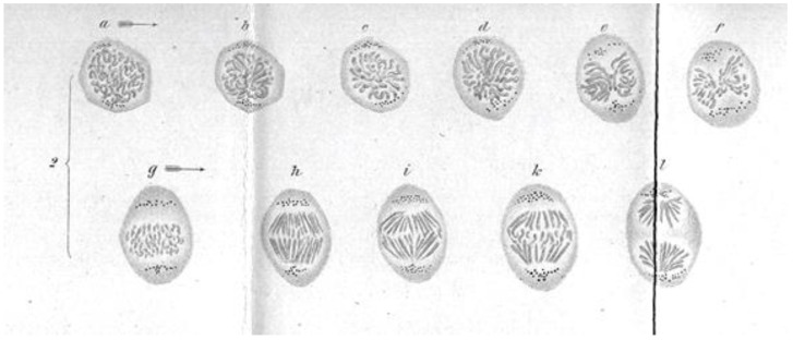
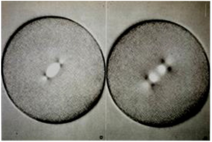
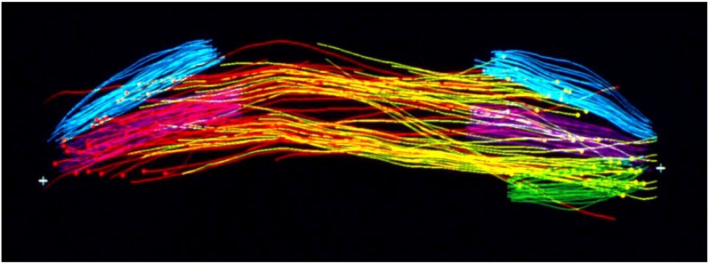
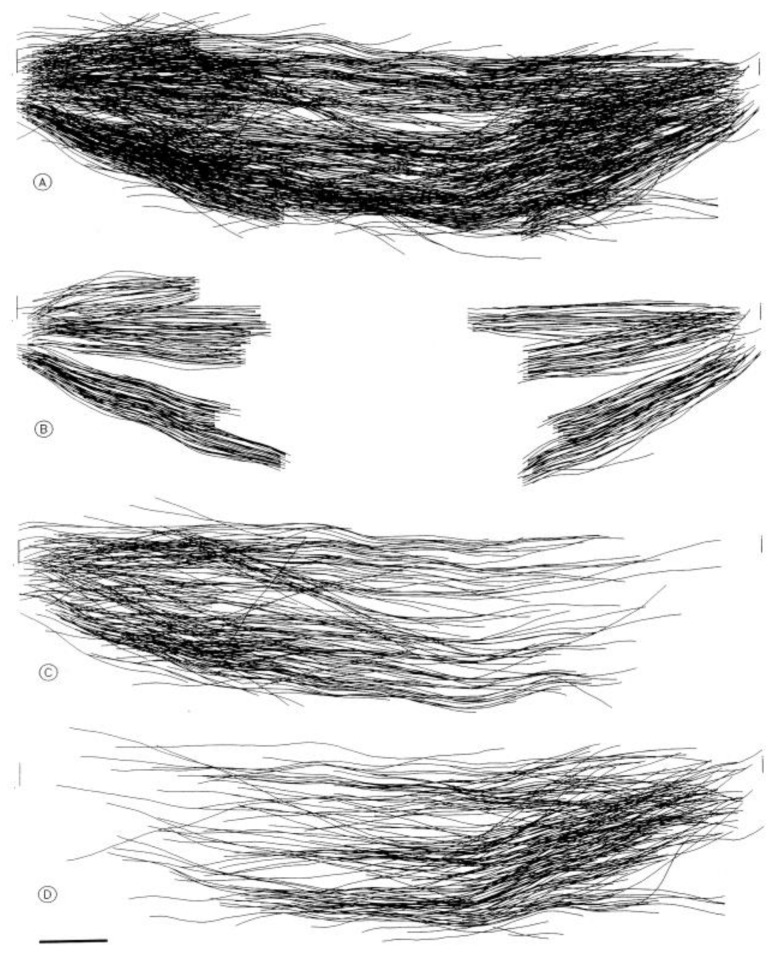
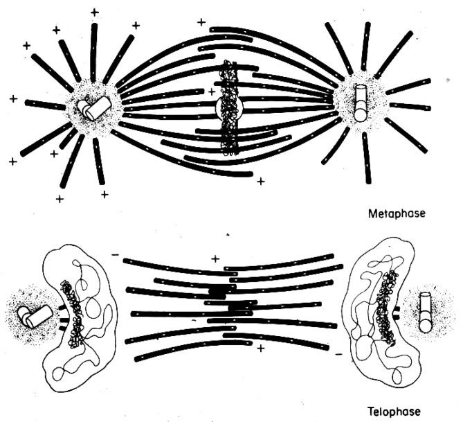
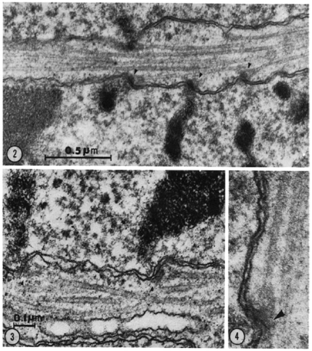
This chapter describes in summary form some of the most important research on chromosome segregation, from the discovery and naming of mitosis in the nineteenth century until around 1990. It gives both historical and scientific background for the nine chapters that follow, each of which provides an up-to-date review of a specific aspect of mitotic mechanism. Here, we trace the fruits of each new technology that allowed a deeper understanding of mitosis and its underlying mechanisms. We describe how light microscopy, including phase, polarization, and fluorescence optics, provided descriptive information about mitotic events and also enabled important experimentation on mitotic functions, such as the dynamics of spindle fibers and the forces generated for chromosome movement. We describe studies by electron microscopy, including quantitative work with serial section reconstructions. We review early results from spindle biochemistry and genetics, coupled to molecular biology, as these methods allowed scholars to identify key molecular components of mitotic mechanisms. We also review hypotheses about mitotic mechanisms whose testing led to a deeper understanding of this fundamental biological event. Our goal is to provide modern scientists with an appreciation of the work that has laid the foundations for their current work and interests.

摘要

本章以总结的形式描述了一些关于染色体分离的最重要研究,从19世纪有丝分裂的发现和命名一直到1990年左右。它为接下来的九章提供了历史和科学背景,每一章都对有丝分裂机制的一个特定方面进行了最新综述。在这里,我们追溯了每项新技术所带来的成果,这些成果使人们对有丝分裂及其潜在机制有了更深入的理解。我们描述了光学显微镜,包括相差、偏振和荧光光学显微镜,如何提供有关有丝分裂事件的描述性信息,以及如何开展关于有丝分裂功能的重要实验,如纺锤体纤维的动态变化以及染色体移动所产生的力。我们描述了电子显微镜的研究,包括对连续切片重建的定量工作。我们回顾了纺锤体生物化学和遗传学与分子生物学相结合的早期研究结果,因为这些方法使学者们能够识别有丝分裂机制的关键分子成分。我们还回顾了关于有丝分裂机制的假说,对这些假说的验证加深了我们对这一基本生物学事件的理解。我们的目标是让现代科学家了解为他们当前的工作和兴趣奠定基础的研究工作。

https://cdn.ncbi.nlm.nih.gov/pmc/blobs/b1bc/5192435/60de2ccbc1b3/biology-05-00055-g021.jpg
https://cdn.ncbi.nlm.nih.gov/pmc/blobs/b1bc/5192435/8dec433e0306/biology-05-00055-g001.jpg
https://cdn.ncbi.nlm.nih.gov/pmc/blobs/b1bc/5192435/e716bec6703e/biology-05-00055-g002.jpg
https://cdn.ncbi.nlm.nih.gov/pmc/blobs/b1bc/5192435/ebfbb95e1e2b/biology-05-00055-g003.jpg
https://cdn.ncbi.nlm.nih.gov/pmc/blobs/b1bc/5192435/e0c2109dc2b9/biology-05-00055-g004.jpg
https://cdn.ncbi.nlm.nih.gov/pmc/blobs/b1bc/5192435/6f0eb4aa64f0/biology-05-00055-g005.jpg
https://cdn.ncbi.nlm.nih.gov/pmc/blobs/b1bc/5192435/2c44ad9d4600/biology-05-00055-g006.jpg
https://cdn.ncbi.nlm.nih.gov/pmc/blobs/b1bc/5192435/92803bb72cc8/biology-05-00055-g007.jpg
https://cdn.ncbi.nlm.nih.gov/pmc/blobs/b1bc/5192435/2d48f47a53e0/biology-05-00055-g008.jpg
https://cdn.ncbi.nlm.nih.gov/pmc/blobs/b1bc/5192435/572ddfedc2e4/biology-05-00055-g009.jpg
https://cdn.ncbi.nlm.nih.gov/pmc/blobs/b1bc/5192435/a1f48130dc2f/biology-05-00055-g010.jpg
https://cdn.ncbi.nlm.nih.gov/pmc/blobs/b1bc/5192435/50d43119d7f4/biology-05-00055-g011.jpg
https://cdn.ncbi.nlm.nih.gov/pmc/blobs/b1bc/5192435/85ee024d5acc/biology-05-00055-g012.jpg
https://cdn.ncbi.nlm.nih.gov/pmc/blobs/b1bc/5192435/741068c853fc/biology-05-00055-g013.jpg
https://cdn.ncbi.nlm.nih.gov/pmc/blobs/b1bc/5192435/4f59288bdcf0/biology-05-00055-g014.jpg
https://cdn.ncbi.nlm.nih.gov/pmc/blobs/b1bc/5192435/74ba9a5fe4f8/biology-05-00055-g015.jpg
https://cdn.ncbi.nlm.nih.gov/pmc/blobs/b1bc/5192435/76f4834ddbf9/biology-05-00055-g016.jpg
https://cdn.ncbi.nlm.nih.gov/pmc/blobs/b1bc/5192435/ab7e9edb8874/biology-05-00055-g017.jpg
https://cdn.ncbi.nlm.nih.gov/pmc/blobs/b1bc/5192435/3ae010d8972b/biology-05-00055-g018.jpg
https://cdn.ncbi.nlm.nih.gov/pmc/blobs/b1bc/5192435/78b402c9dd6b/biology-05-00055-g019.jpg
https://cdn.ncbi.nlm.nih.gov/pmc/blobs/b1bc/5192435/f6ffa62ca295/biology-05-00055-g020.jpg
https://cdn.ncbi.nlm.nih.gov/pmc/blobs/b1bc/5192435/60de2ccbc1b3/biology-05-00055-g021.jpg

相似文献

1
A Brief History of Research on Mitotic Mechanisms.有丝分裂机制研究简史
Biology (Basel). 2016 Dec 21;5(4):55. doi: 10.3390/biology5040055.
2
Mechanisms of chromosome biorientation and bipolar spindle assembly analyzed by computational modeling.通过计算建模分析染色体的双定向和双极纺锤体组装的机制。
Elife. 2020 Feb 13;9:e48787. doi: 10.7554/eLife.48787.
3
Measurement of Microtubule Half-Life and Poleward Flux in the Mitotic Spindle by Photoactivation of Fluorescent Tubulin.通过荧光微管蛋白的光激活测量有丝分裂纺锤体中的微管半衰期和极向流。
Methods Mol Biol. 2020;2101:235-246. doi: 10.1007/978-1-0716-0219-5_15.
4
Assessing the Contributions of Motor Enzymes and Microtubule Dynamics to Mitotic Chromosome Motions.评估运动酶和微管动力学对有丝分裂染色体运动的贡献。
Annu Rev Cell Dev Biol. 2017 Oct 6;33:1-22. doi: 10.1146/annurev-cellbio-100616-060827.
5
Centromere Dysfunction Compromises Mitotic Spindle Pole Integrity.着丝粒功能障碍破坏有丝分裂纺锤体极的完整性。
Curr Biol. 2019 Sep 23;29(18):3072-3080.e5. doi: 10.1016/j.cub.2019.07.052. Epub 2019 Sep 5.
6
Mitotic spindle assembly and chromosome segregation: refocusing on microtubule dynamics.有丝分裂纺锤体组装与染色体分离:重新聚焦于微管动力学
Mol Cell. 2004 Aug 13;15(3):317-27. doi: 10.1016/j.molcel.2004.07.012.
7
Modelling chromosome dynamics in mitosis: a historical perspective on models of metaphase and anaphase in eukaryotic cells.有丝分裂中染色体动力学建模:真核细胞中期和后期模型的历史视角
Interface Focus. 2014 Jun 6;4(3):20130073. doi: 10.1098/rsfs.2013.0073.
8
Centrosomes and the art of mitotic spindle maintenance.中心体与有丝分裂纺锤体的维护艺术。
Int Rev Cell Mol Biol. 2014;313:179-217. doi: 10.1016/B978-0-12-800177-6.00006-2.
9
More than two populations of microtubules comprise the dynamic mitotic spindle.有超过两种微管群体构成了有丝分裂纺锤体的动态结构。
J Cell Sci. 2022 Feb 1;135(3). doi: 10.1242/jcs.258745. Epub 2022 Feb 2.
10
Mechanisms of mitotic spindle assembly and function.有丝分裂纺锤体组装与功能的机制。
Int Rev Cytol. 2008;265:111-58. doi: 10.1016/S0074-7696(07)65003-7.

引用本文的文献

1
Insights into the role of phosphorylation on microtubule cross-linking by PRC1.对PRC1磷酸化在微管交联中作用的见解。
Mol Biol Cell. 2025 Mar 1;36(3):ar34. doi: 10.1091/mbc.E24-12-0565. Epub 2025 Jan 22.
2
Force generation and resistance in human mitosis.人类有丝分裂中的力产生与阻力
Biophys Rev. 2024 Sep 28;16(5):551-562. doi: 10.1007/s12551-024-01235-0. eCollection 2024 Oct.
3
Microtubule choreography: spindle self-organization during cell division.微管编排:细胞分裂过程中的纺锤体自我组织

本文引用的文献

1
[Not Available].[无可用内容]
Wilhelm Roux Arch Entwickl Mech Org. 1929 Jun;118(1):359-484. doi: 10.1007/BF02108879.
2
Anaphase B.后期B
Biology (Basel). 2016 Dec 8;5(4):51. doi: 10.3390/biology5040051.
3
Bulk cytoplasmic actin and its functions in meiosis and mitosis.细胞质肌动蛋白及其在减数分裂和有丝分裂中的功能。
Biophys Rev. 2024 Sep 30;16(5):613-624. doi: 10.1007/s12551-024-01236-z. eCollection 2024 Oct.
4
New characterization and safety evaluation of human limbal stem cells used in clinical application: fidelity of mitotic process and mitotic spindle morphologies.临床应用中人角膜缘干细胞的新特征和安全性评估:有丝分裂过程和有丝分裂纺锤体形态的保真度。
Stem Cell Res Ther. 2023 Dec 13;14(1):368. doi: 10.1186/s13287-023-03586-z.
5
Label-Free Cytometric Evaluation of Mitosis via Stimulated Raman Scattering Microscopy and Spectral Phasor Analysis.无标记流式细胞术通过受激拉曼散射显微镜和光谱相分析评估有丝分裂
Anal Chem. 2023 May 9;95(18):7244-7253. doi: 10.1021/acs.analchem.3c00212. Epub 2023 Apr 25.
6
Coupling of microtubule bundles isolates them from local disruptions to set the structural stability of the anaphase spindle.微管束的耦合将其与局部破坏隔离开来,从而确定了后期纺锤体的结构稳定性。
Proc Natl Acad Sci U S A. 2022 Sep 27;119(39):e2204068119. doi: 10.1073/pnas.2204068119. Epub 2022 Sep 19.
7
Mitotic H3K9ac is controlled by phase-specific activity of HDAC2, HDAC3, and SIRT1.有丝分裂期 H3K9ac 的水平受 HDAC2、HDAC3 和 SIRT1 的相分离活性控制。
Life Sci Alliance. 2022 Aug 18;5(10). doi: 10.26508/lsa.202201433. Print 2022 Oct.
8
Directly Measuring Forces within Reconstituted Active Microtubule Bundles.直接测量重建活性微管束内的力。
J Vis Exp. 2022 May 10(183). doi: 10.3791/63819.
9
Fifteen compelling open questions in plant cell biology.植物细胞生物学十五个极富挑战性的开放性问题。
Plant Cell. 2022 Jan 20;34(1):72-102. doi: 10.1093/plcell/koab225.
10
Shake It Off: The Elimination of Erroneous Kinetochore-Microtubule Attachments and Chromosome Oscillation.《摇掉它:错误的动粒-微管连接的消除和染色体震荡》。
Int J Mol Sci. 2021 Mar 20;22(6):3174. doi: 10.3390/ijms22063174.
Curr Biol. 2011 Oct 11;21(19):R825-30. doi: 10.1016/j.cub.2011.07.043.
4
Equilibrium of trivalents and the mechanism of chromosome movements.三价体的平衡与染色体运动机制
Hereditas. 1945;31(3-4):498.
5
Hydrostatic pressure effects upon the spindle figure and chromosome movement; experiments on the meiotic divisions of Trade-scantia pollen mother cells.流体静压力对纺锤体形态和染色体移动的影响;关于紫露草花粉母细胞减数分裂的实验
Biol Bull. 1946 Oct;91(02):145-69.
6
The distribution of spindle microtubules during mitosis in cultured human cells.有丝分裂过程中培养的人细胞纺锤体微管的分布。
J Cell Biol. 1971 May 1;49(2):468-97. doi: 10.1083/jcb.49.2.468.
7
CONTRIBUTIONS TO THE KNOWLEDGE OF THE CELL AND ITS VITAL PROCESSES.对细胞及其生命过程知识的贡献
J Cell Biol. 1965 Apr 1;25(1):3-69.
8
Computer simulations predict that chromosome movements and rotations accelerate mitotic spindle assembly without compromising accuracy.计算机模拟预测,染色体的移动和旋转可加速有丝分裂纺锤体组装,且不影响准确性。
Proc Natl Acad Sci U S A. 2009 Sep 15;106(37):15708-13. doi: 10.1073/pnas.0908261106. Epub 2009 Aug 26.
9
AN APPARENT CASE OF MONOCENTRIC MITOSIS IN SCIARA (DIPTERA).果蝇(双翅目)单中心有丝分裂的一个明显案例。
Science. 1926 Feb 12;63(1624):190-1. doi: 10.1126/science.63.1624.190.
10
Dynein: A Protein with Adenosine Triphosphatase Activity from Cilia.动力蛋白:纤毛中的一种具有三磷酸腺苷酶活性的蛋白质。
Science. 1965 Jul 23;149(3682):424-6. doi: 10.1126/science.149.3682.424.