Athmanathan Sreedharan, Reddy Sesha B, Nutheti Rishita, Rao Gullapalli N
Jhaveri Microbiology Center, Prof. Brien Holden Eye Research Center, Hyderabad Eye Research Foundation, L. V. Prasad Eye Institute, L. V. Prasad Marg, Banjara Hills, Hyderabad, India.
BMC Ophthalmol. 2002 Apr 30;2:3. doi: 10.1186/1471-2415-2-3.
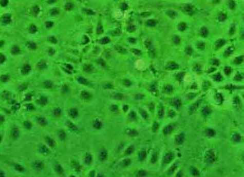
Herpes simplex keratitis (HSK) is a sight threatening ocular infection often requiring a specific and prompt laboratory diagnosis. Isolation of Herpes simplex virus (HSV-1) in culture provides the most reliable and specific method and is considered as the "Gold Standard" in the laboratory diagnosis of HSK in spite of its low sensitivity. Using "cell lines of corneal origin" for virus isolation may be beneficial under such circumstances, since these cells have been shown to be excellent substrates for the growth of HSV-1 isolated from the cornea. We report a comparative study of a novel human corneal epithelial cell line (HCE) and the Vero cell line in the isolation of HSV-1 from corneal scrapings employing a shell vial assay.
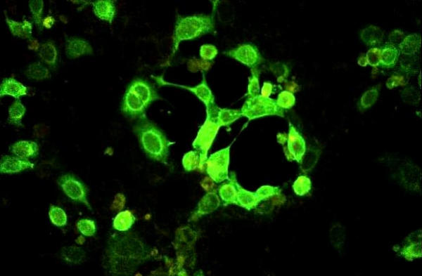
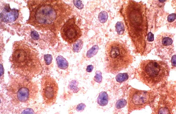
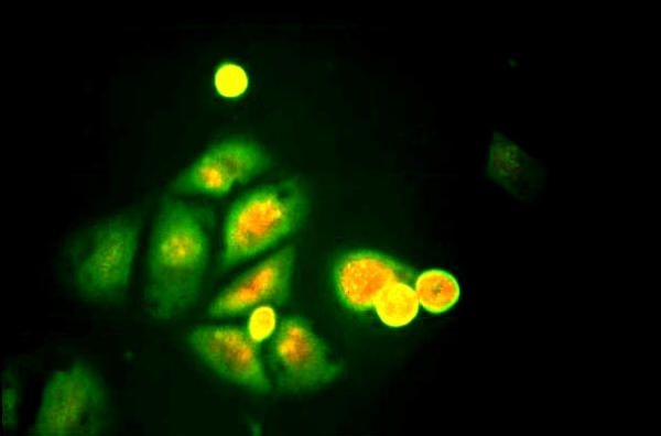
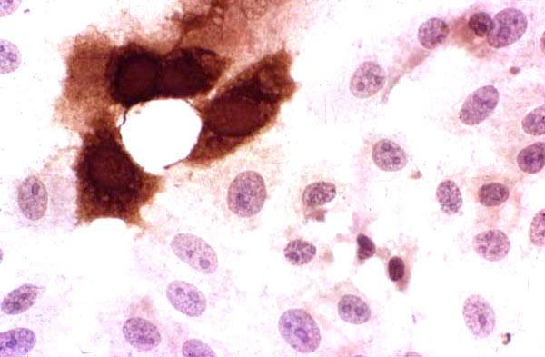
Corneal scrapings were obtained from 17 patients with a clinical diagnosis of HSK. All the cases were confirmed by virological investigations (PCR and viral antigen detection positive, n = 15, PCR positive, n = 1, Viral antigen positive, n = 1). Scrapings obtained from 10 patients with infectious keratitis of non-viral origin were included as controls. All the scrapings were simultaneously inoculated into shell vials of HCE and Vero cells. Cultures were terminated at 24 h post-infection. Isolation of HSV-1 was confirmed using an indirect immunofluorescence/ immunoperoxidase assay.
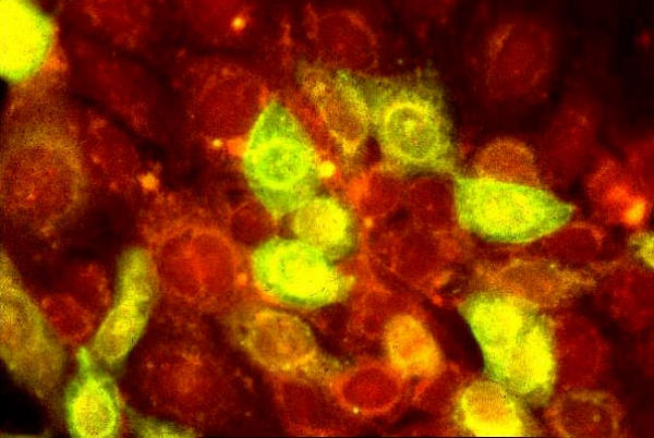
Virus could be isolated using both or either of the cell lines in 10/17 (58.82%) patients with HSK. HSV-1 was isolated from 10/17 (58.82%) and 4/17(23.52%) specimens in HCE and Vero cells, respectively (P = 0.036). None of the controls yielded HSV-1. While all the 10 (100%) strains were isolated in HCE, Vero yielded only 4/10 (40%) strains in the shell vial culture (P = 0.014).
HCE showed a statistically significant difference in the virus isolation rate with respect to Vero cells. HCE may be an excellent alternative cell line for the isolation of HSV-1, especially from corneal scrapings, for the laboratory diagnosis of HSK.
单纯疱疹病毒性角膜炎(HSK)是一种威胁视力的眼部感染,通常需要进行特定且快速的实验室诊断。尽管其敏感性较低,但在培养物中分离单纯疱疹病毒(HSV - 1)是最可靠且特异的方法,被视为HSK实验室诊断的“金标准”。在这种情况下,使用“角膜来源的细胞系”进行病毒分离可能是有益的,因为这些细胞已被证明是从角膜分离出的HSV - 1生长的优良底物。我们报告了一项关于新型人角膜上皮细胞系(HCE)和Vero细胞系在采用空斑试验从角膜刮片中分离HSV - 1的比较研究。
从17例临床诊断为HSK的患者获取角膜刮片。所有病例均经病毒学检查确诊(PCR和病毒抗原检测阳性,n = 15;PCR阳性,n = 1;病毒抗原阳性,n = 1)。将从10例非病毒源性感染性角膜炎患者获取的刮片作为对照。所有刮片同时接种到HCE和Vero细胞的空斑管中。感染后24小时终止培养。使用间接免疫荧光/免疫过氧化物酶测定法确认HSV - 1的分离。
在17例HSK患者中,10例(58.82%)患者使用两种细胞系中的一种或两种均可分离出病毒。在HCE和Vero细胞中,分别从17份标本中的10份(58.82%)和4份(23.52%)分离出HSV - 1(P = 0.036)。所有对照均未分离出HSV - 1。在空斑管培养中,所有10株(100%)菌株在HCE中被分离出来,而Vero仅分离出4/10(40%)菌株(P = 0.014)。
HCE在病毒分离率方面与Vero细胞相比具有统计学显著差异。HCE可能是用于分离HSV - 1的优良替代细胞系,特别是用于从角膜刮片中分离HSV - 1,以进行HSK的实验室诊断。