Kubicki Marek, Westin Carl-Fredrik, Maier Stephan E, Frumin Melissa, Nestor Paul G, Salisbury Dean F, Kikinis Ron, Jolesz Ferenc A, McCarley Robert W, Shenton Martha E
Department of Psychiatry, Harvard Medical School and VA Boston Healthcare System-Brockton Division, MA 02301, USA.
Am J Psychiatry. 2002 May;159(5):813-20. doi: 10.1176/appi.ajp.159.5.813.
Disruptions in connectivity between the frontal and temporal lobes may explain some of the symptoms observed in schizophrenia. Conventional magnetic resonance imaging (MRI) studies, however, have not shown compelling evidence for white matter abnormalities, because white matter fiber tracts cannot be visualized by conventional MRI. Diffusion tensor imaging is a relatively new technique that can detect subtle white matter abnormalities in vivo by assessing the degree to which directionally organized fibers have lost their normal integrity. The first three diffusion tensor imaging studies in schizophrenia showed lower anisotropic diffusion, relative to comparison subjects, in whole-brain white matter, prefrontal and temporal white matter, and the corpus callosum, respectively. Here the authors focus on fiber tracts forming temporal-frontal connections.
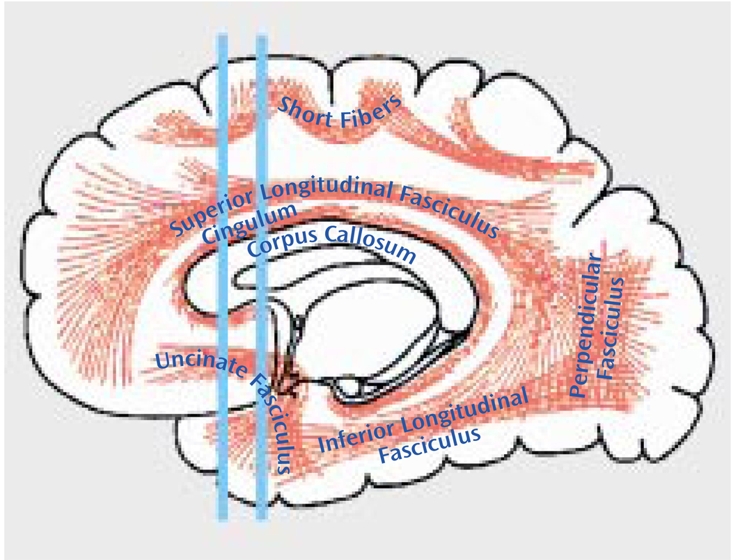
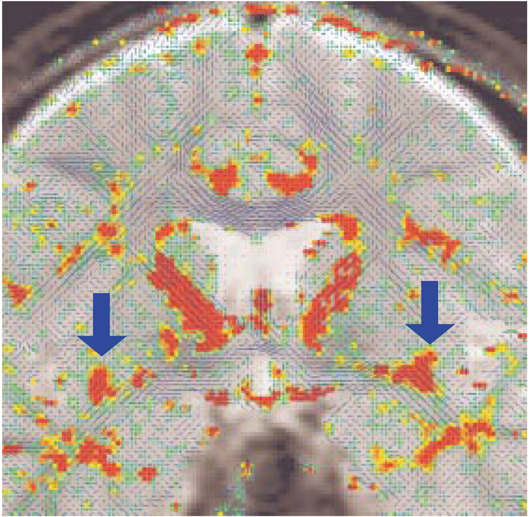
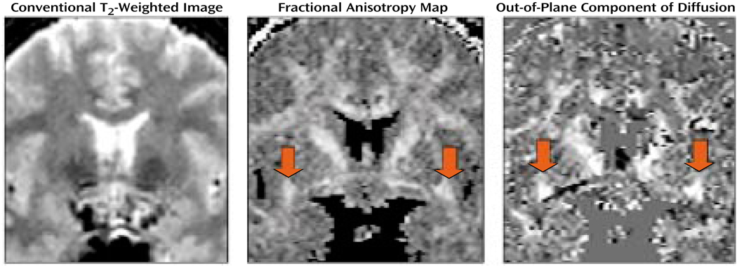
Anisotropic diffusion was assessed in the uncinate fasciculus, the most prominent white matter tract connecting temporal and frontal brain regions, in 15 patients with chronic schizophrenia and 18 normal comparison subjects. A 1.5-T GE Echospeed system was used to acquire 4-mm-thick coronal line-scan diffusion tensor images. Maps of the fractional anisotropy were generated to quantify the water diffusion within the uncinate fasciculus.
Findings revealed a group-by-side interaction for fractional anisotropy and for uncinate fasciculus area, derived from automatic segmentation. The patients with schizophrenia showed a lack of normal left-greater-than-right asymmetry seen in the comparison subjects.
These findings demonstrate the importance of investigating white matter tracts in vivo in schizophrenia and support the hypothesis of a disruption in the normal pattern of connectivity between temporal and frontal brain regions in schizophrenia.
额叶与颞叶之间连接的中断可能解释精神分裂症中观察到的一些症状。然而,传统的磁共振成像(MRI)研究并未显示出白质异常的有力证据,因为传统MRI无法显示白质纤维束。扩散张量成像(DTI)是一种相对较新的技术,它可以通过评估定向排列的纤维丧失其正常完整性的程度,在活体中检测细微的白质异常。最初三项关于精神分裂症的扩散张量成像研究分别显示,与对照组相比,全脑白质、前额叶和颞叶白质以及胼胝体的各向异性扩散较低。本文作者聚焦于形成颞叶 - 额叶连接的纤维束。
在15例慢性精神分裂症患者和18名正常对照者中,评估了钩束(连接颞叶和额叶脑区的最显著白质束)的各向异性扩散。使用1.5-T的GE Echospeed系统获取4毫米厚的冠状线扫描扩散张量图像。生成分数各向异性图以量化钩束内的水扩散。
研究结果揭示了分数各向异性和通过自动分割得出的钩束面积的组间交互作用。精神分裂症患者缺乏在对照者中可见的正常的左大于右的不对称性。
这些发现证明了在活体中研究精神分裂症白质束的重要性,并支持精神分裂症中颞叶和额叶脑区之间正常连接模式中断的假说。