Prior Ian A, Muncke Cornelia, Parton Robert G, Hancock John F
Department of Pathology and Institute for Molecular Bioscience, University of Queensland, Brisbane, Queensland 4006, Australia.
J Cell Biol. 2003 Jan 20;160(2):165-70. doi: 10.1083/jcb.200209091. Epub 2003 Jan 13.
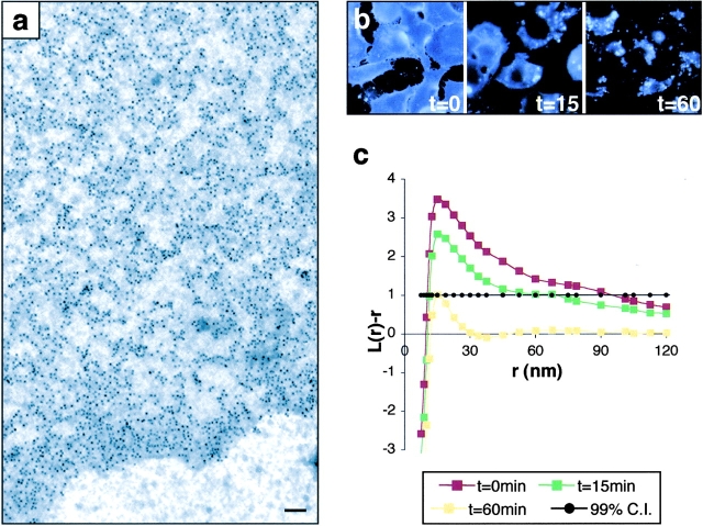
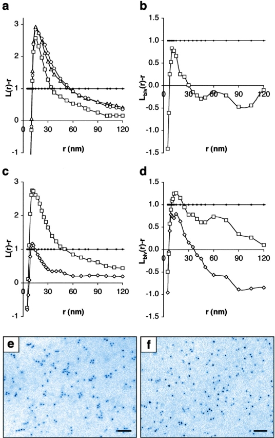
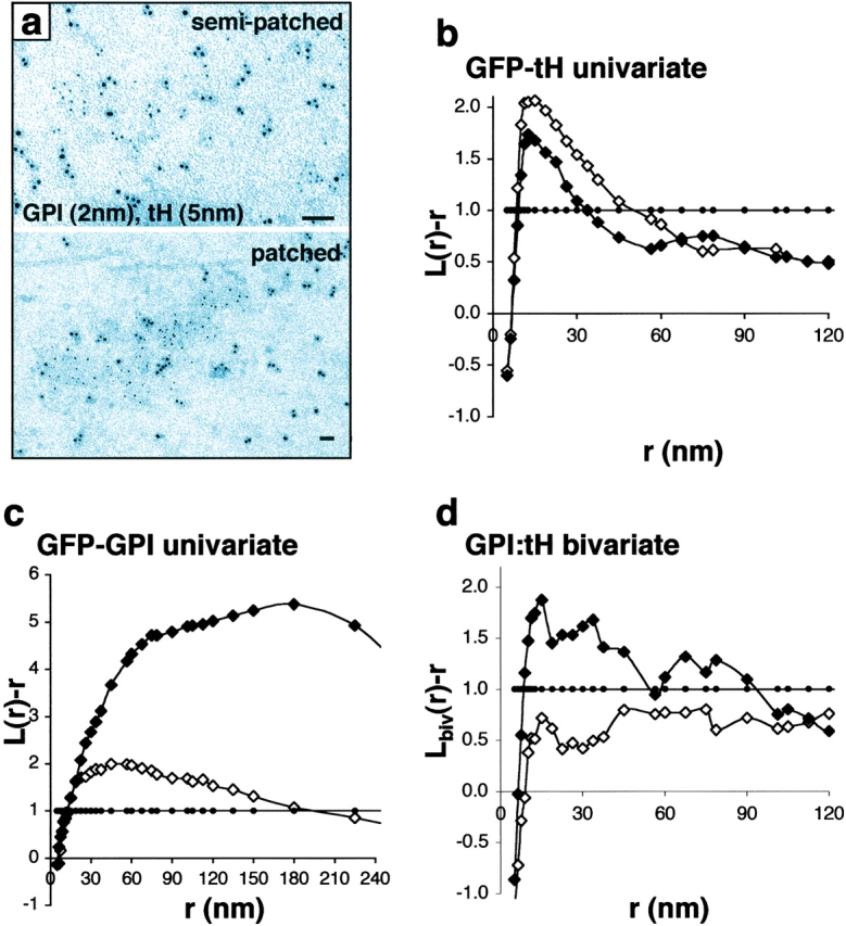
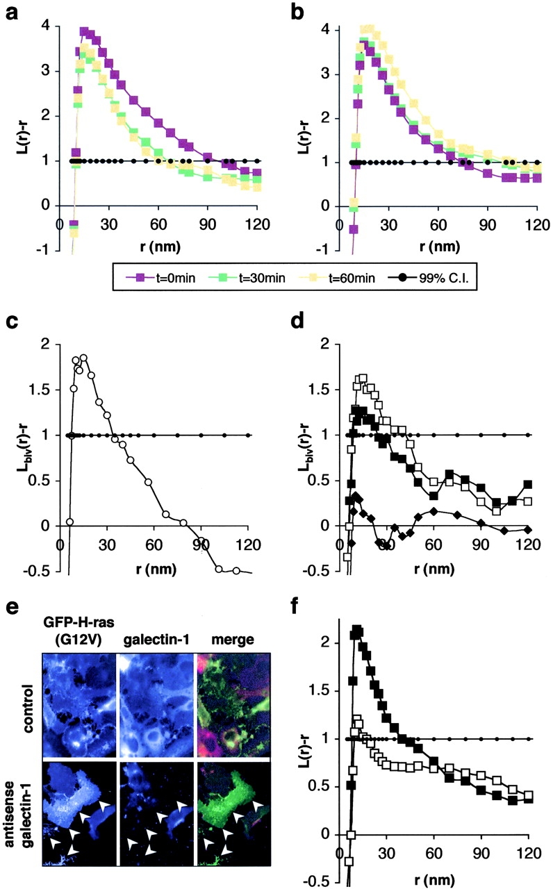
Localization of signaling complexes to specific microdomains coordinates signal transduction at the plasma membrane. Using immunogold electron microscopy of plasma membrane sheets coupled with spatial point pattern analysis, we have visualized morphologically featureless microdomains, including lipid rafts, in situ and at high resolution. We find that an inner-plasma membrane lipid raft marker displays cholesterol-dependent clustering in microdomains with a mean diameter of 44 nm that occupy 35% of the cell surface. Cross-linking an outer-leaflet raft protein results in the redistribution of inner leaflet rafts, but they retain their modular structure. Analysis of Ras microlocalization shows that inactive H-ras is distributed between lipid rafts and a cholesterol-independent microdomain. Conversely, activated H-ras and K-ras reside predominantly in nonoverlapping, cholesterol-independent microdomains. Galectin-1 stabilizes the association of activated H-ras with these nonraft microdomains, whereas K-ras clustering is supported by farnesylation, but not geranylgeranylation. These results illustrate that the inner plasma membrane comprises a complex mosaic of discrete microdomains. Differential spatial localization within this framework can likely account for the distinct signal outputs from the highly homologous Ras proteins.
信号复合物定位于特定微结构域可协调质膜上的信号转导。通过结合空间点模式分析的质膜片免疫金电子显微镜技术,我们已在原位高分辨率观察到包括脂筏在内的无形态特征的微结构域。我们发现,一种质膜内层脂筏标记物在平均直径为44 nm、占据细胞表面35%的微结构域中呈现胆固醇依赖性聚集。交联外层脂筏蛋白会导致内层脂筏重新分布,但它们仍保留其模块化结构。对Ras微定位的分析表明,无活性的H-Ras分布于脂筏和一个不依赖胆固醇的微结构域之间。相反,活化的H-Ras和K-Ras主要位于不重叠的、不依赖胆固醇的微结构域中。半乳糖凝集素-1稳定活化的H-Ras与这些非脂筏微结构域的结合,而K-Ras的聚集由法尼基化而非香叶基香叶基化支持。这些结果表明,质膜内层由离散微结构域的复杂镶嵌组成。在此框架内的差异空间定位可能解释了高度同源的Ras蛋白产生的不同信号输出。