Grabowski P, Kühnel T, Mühr-Wilkenshoff F, Heine B, Stein H, Höpfner M, Germer C T, Scherübl H
Medical Clinic I, Gastroenterology/Infectious Diseases/Rheumatology, Benjamin Franklin Clinics, FU Berlin, Hindenburgdamm 30, 12200 Berlin, Germany.
Br J Cancer. 2003 Jan 13;88(1):115-9. doi: 10.1038/sj.bjc.6600696.
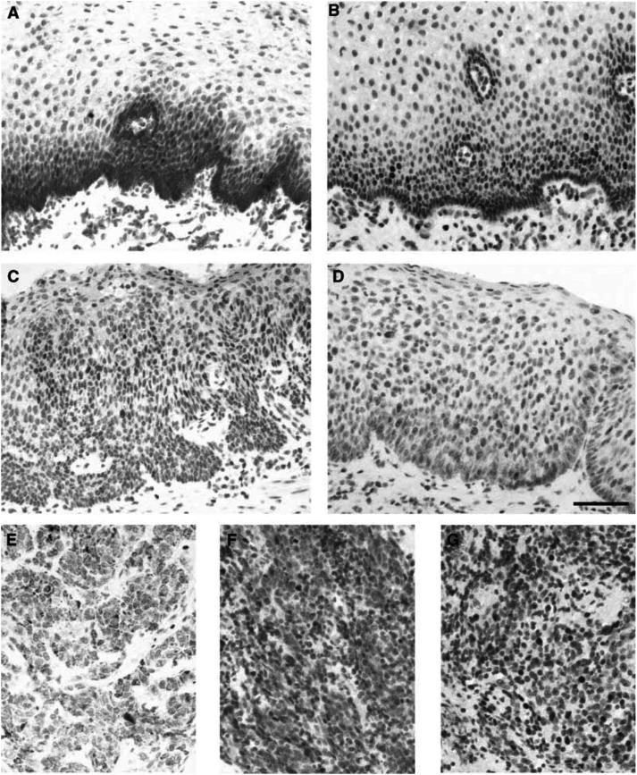
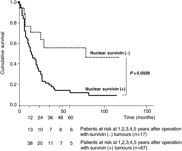
Survivin, a new member of the family of apoptosis inhibitors, is expressed almost exclusively in proliferating cells, above all in cancers. Subcellular localisation and prognostic implications of the survivin protein have not yet been determined in oesophageal squamous cell carcinoma. The survival of 84 patients with oesophageal squamous cell carcinomas was correlated with the extent of immunohistochemical survivin expression in tumour cell nuclei. Tumours were scored positive when >5% cells stained positive. Patients were followed up for at least 5 years or until death. In normal oesophageal squamous cell epithelium, some cytoplasmic survivin expression was detected in the basal cells, whereas proliferating cells showed nuclear staining of survivin. Nuclear expression of survivin was also detected in 67 cancers (80%). The mean survival for patients of this group (28 months, range 20-36) was significantly less than that for patients without survivin expression in the tumour cell nuclei (108 months, range 62-154, P=0.003). Using univariate analysis, nuclear survivin expression (P=0.003), tumour depth (P=0.001), lymph node metastasis (P=0.003) and stage (P<0.001) were the best predictors of survival. In contrast, cytoplasmic survivin staining was noted in 53 (63%) tumours and had no prognostic relevance. In conclusion, the analysis of nuclear survivin expression identifies subgroups in oesophageal squamous cell cancer with favourable (survivin(-)) or with poor prognosis (survivin(+)). We suggest that the determination of nuclear survivin expression could be used to individualise therapeutic strategies in oesophageal squamous cell cancer in the future.
存活素是凋亡抑制蛋白家族的新成员,几乎仅在增殖细胞中表达,尤其是在癌细胞中。食管鳞状细胞癌中存活素蛋白的亚细胞定位及其预后意义尚未明确。对84例食管鳞状细胞癌患者的生存情况与肿瘤细胞核中免疫组化检测的存活素表达程度进行相关性分析。当>5%的细胞染色呈阳性时,肿瘤被判定为阳性。对患者进行至少5年的随访或直至死亡。在正常食管鳞状上皮细胞中,仅在基底细胞中检测到一些细胞质存活素表达,而增殖细胞显示存活素的细胞核染色。在67例癌症(80%)中也检测到存活素的细胞核表达。该组患者的平均生存期(28个月,范围20 - 36个月)显著短于肿瘤细胞核中无存活素表达的患者(108个月,范围62 - 154个月,P = 0.003)。单因素分析显示,细胞核存活素表达(P = 0.003)、肿瘤深度(P = 0.001)、淋巴结转移(P = 0.003)和分期(P < 0.001)是生存的最佳预测指标。相比之下,53例(63%)肿瘤中观察到细胞质存活素染色,其无预后相关性。总之,对细胞核存活素表达的分析可识别食管鳞状细胞癌中预后良好(存活素阴性)或预后不良(存活素阳性)的亚组。我们建议,测定细胞核存活素表达未来可用于食管鳞状细胞癌治疗策略的个体化。