Waterman-Storer C M, Salmon E D
Department of Biology, 607 Fordham Hall, University of North Carolina, Chapel Hill, North Carolina 27599-3280, USA.
J Cell Biol. 1997 Oct 20;139(2):417-34. doi: 10.1083/jcb.139.2.417.
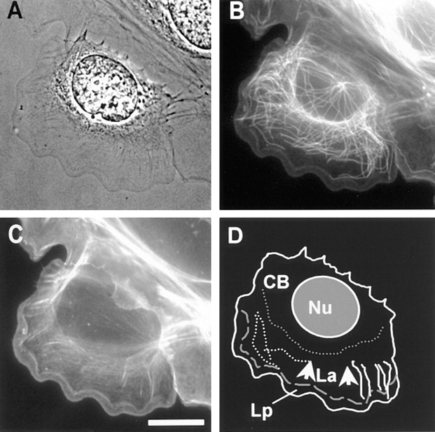
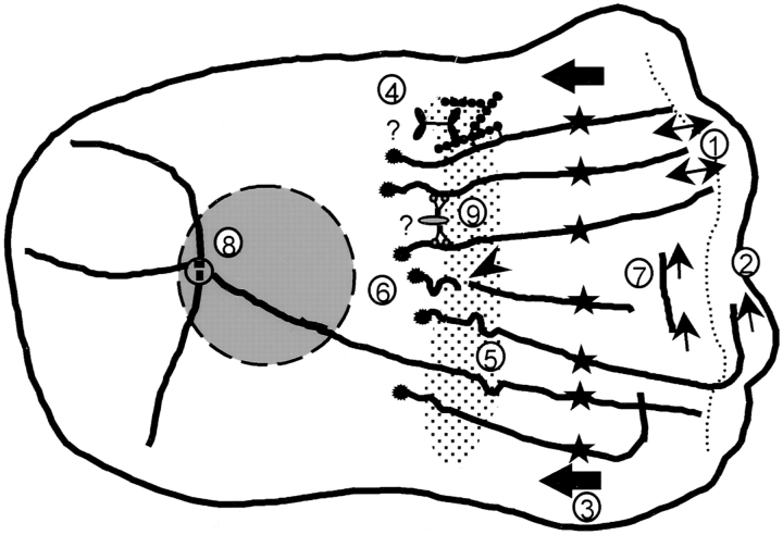
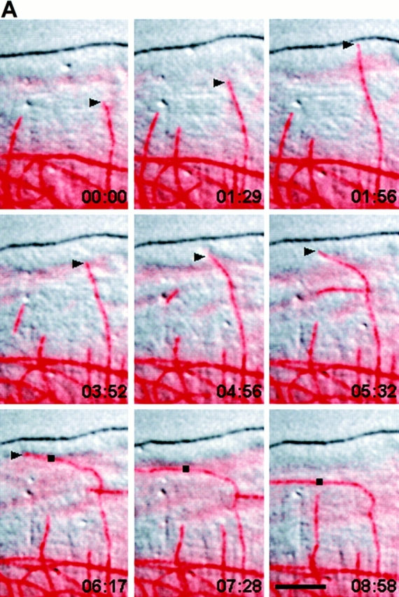
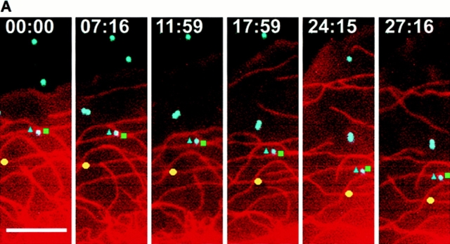
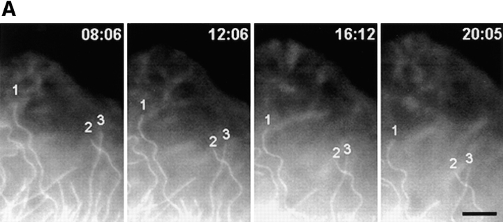
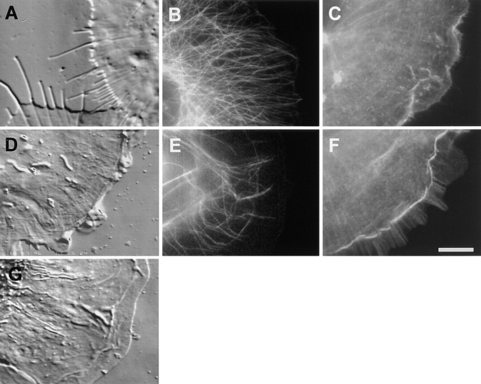
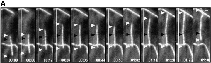
We have discovered several novel features exhibited by microtubules (MTs) in migrating newt lung epithelial cells by time-lapse imaging of fluorescently labeled, microinjected tubulin. These cells exhibit leading edge ruffling and retrograde flow in the lamella and lamellipodia. The plus ends of lamella MTs persist in growth perpendicular to the leading edge until they reach the base of the lamellipodium, where they oscillate between short phases of growth and shortening. Occasionally "pioneering" MTs grow into the lamellipodium, where microtubule bending and reorientation parallel to the leading edge is associated with retrograde flow. MTs parallel to the leading edge exhibit significantly different dynamics from MTs perpendicular to the cell edge. Both parallel MTs and photoactivated fluorescent marks on perpendicular MTs move rearward at the 0.4 mircon/min rate of retrograde flow in the lamella. MT rearward transport persists when MT dynamic instability is inhibited by 100-nM nocodazole but is blocked by inhibition of actomyosin by cytochalasin D or 2,3-butanedione-2-monoxime. Rearward flow appears to cause MT buckling and breaking in the lamella. 80% of free minus ends produced by breakage are stable; the others shorten and pause, leading to MT treadmilling. Free minus ends of unknown origin also depolymerize into the field of view at the lamella. Analysis of MT dynamics at the centrosome shows that these minus ends do not arise by centrosomal ejection and that approximately 80% of the MTs in the lamella are not centrosome bound. We propose that actomyosin-based retrograde flow of MTs causes MT breakage, forming quasi-stable noncentrosomal MTs whose turnover is regulated primarily at their minus ends.
通过对荧光标记、显微注射的微管蛋白进行延时成像,我们发现了蝾螈肺上皮细胞迁移过程中微管(MTs)呈现的几个新特征。这些细胞在片状伪足和丝状伪足中表现出前沿褶皱和逆向流动。片状微管的正端持续垂直于前沿生长,直到到达丝状伪足的基部,在那里它们在短时间的生长和缩短阶段之间振荡。偶尔会有“先锋”微管生长到丝状伪足中,在那里微管弯曲并与前沿平行重新定向与逆向流动相关。与前沿平行的微管与垂直于细胞边缘的微管表现出显著不同的动力学。平行微管和垂直微管上的光活化荧光标记都以片状伪足中0.4微米/分钟的逆向流动速率向后移动。当100 nM诺考达唑抑制微管动态不稳定性时,微管向后运输持续存在,但细胞松弛素D或2,3-丁二酮-2-单肟抑制肌动球蛋白会阻断这种运输。向后流动似乎会导致微管在片状伪足中弯曲和断裂。断裂产生的游离负端80%是稳定的;其他的则缩短并暂停,导致微管踏车运动。未知来源的游离负端也在片状伪足处解聚到视野中。对中心体处微管动力学的分析表明,这些负端不是由中心体排出产生的,并且片状伪足中约80%的微管不与中心体结合。我们提出,基于肌动球蛋白的微管逆向流动导致微管断裂,形成准稳定的非中心体微管,其周转主要在负端受到调节。