Rosenfeld S J, Yoshimoto K, Kajigaya S, Anderson S, Young N S, Field A, Warrener P, Bansal G, Collett M S
Cell Biology Section, National Heart, Lung and Blood Institute, Bethesda, Maryland 20817.
J Clin Invest. 1992 Jun;89(6):2023-9. doi: 10.1172/JCI115812.
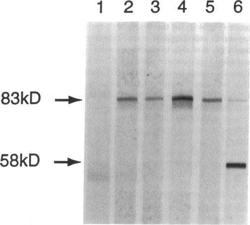
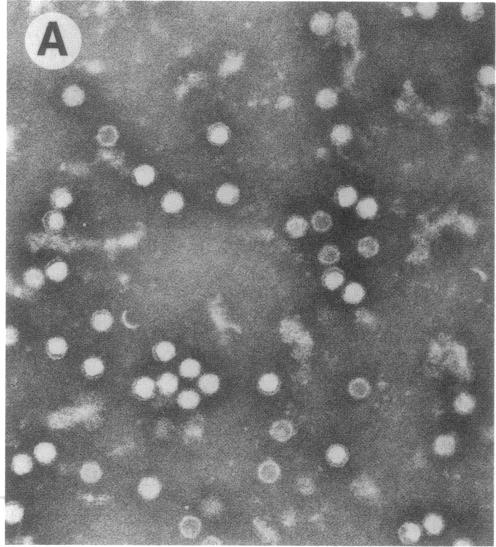
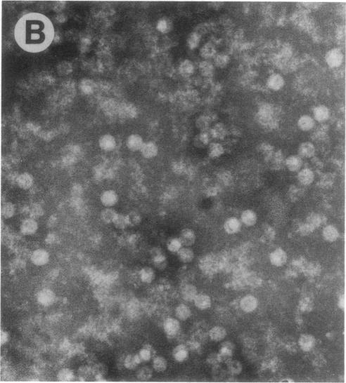
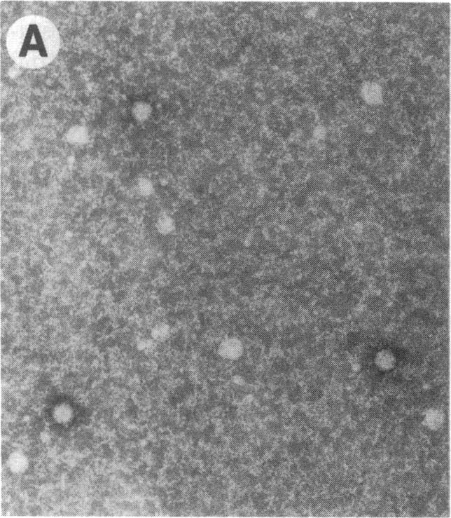
Capsids of the B19 parvovirus are composed of major (VP2; 58 kD) and minor (VP1; 83 kD) structural proteins. These proteins are identical except for a unique 226 amino acid region at the amino terminus of VP1. Previous immunization studies with recombinant empty capsids have demonstrated that the presence of VP1 was required to elicit virus-neutralizing antibody activity. However, to date, neutralizing epitopes have been identified only on VP2. Crystallographic studies of a related parvovirus (canine parvovirus) suggested the unique amino-terminal portion of VP1 assumed an internal position within the viral capsid. To determine the position of VP1 in both empty capsids and virions, we expressed a fusion protein containing the unique region of VP1. Antisera raised to this protein recognized recombinant empty capsids containing VP1 and VP2, but not those containing VP2 alone, in an enzyme-linked immunosorbent assay. The antisera immunoprecipitated both recombinant empty capsids and human plasma-derived virions, and agglutinated the latter as shown by immune electron microscopy. The sera contained potent neutralizing activity for virus infectivity in vitro. These data indicate that a portion of the amino terminus of VP1 is located on the virion surface, and that this region contains intrinsic neutralizing determinants. The external location of the VP1-specific tail may provide a site for engineered heterologous epitope presentation in novel recombinant vaccines.
B19细小病毒的衣壳由主要结构蛋白(VP2;58千道尔顿)和次要结构蛋白(VP1;83千道尔顿)组成。除了VP1氨基末端有一个独特的226个氨基酸的区域外,这些蛋白质是相同的。先前对重组空衣壳的免疫研究表明,VP1的存在是引发病毒中和抗体活性所必需的。然而,迄今为止,仅在VP2上鉴定出中和表位。对一种相关细小病毒(犬细小病毒)的晶体学研究表明,VP1独特的氨基末端部分在病毒衣壳内处于内部位置。为了确定VP1在空衣壳和病毒粒子中的位置,我们表达了一种包含VP1独特区域的融合蛋白。在酶联免疫吸附测定中,针对该蛋白产生的抗血清识别含有VP1和VP2的重组空衣壳,但不识别仅含有VP2的重组空衣壳。抗血清免疫沉淀重组空衣壳和人血浆来源的病毒粒子,并如免疫电子显微镜所示凝集后者。这些血清在体外对病毒感染性具有强大的中和活性。这些数据表明,VP1氨基末端的一部分位于病毒粒子表面,并且该区域含有内在的中和决定簇。VP1特异性尾部的外部位置可能为新型重组疫苗中工程化异源表位的呈现提供一个位点。