Preuss D, Mulholland J, Franzusoff A, Segev N, Botstein D
Department of Biochemistry, Beckman Center, Stanford University, CA 94305.
Mol Biol Cell. 1992 Jul;3(7):789-803. doi: 10.1091/mbc.3.7.789.
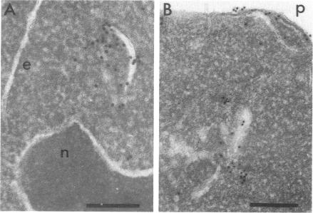
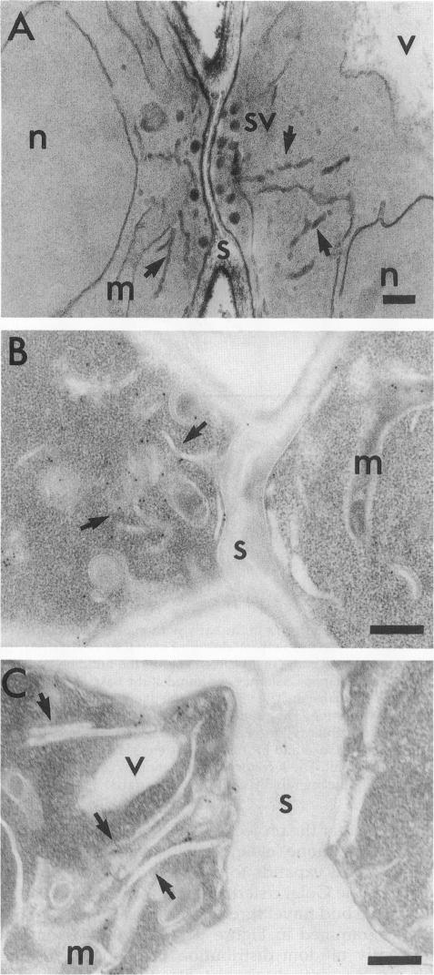
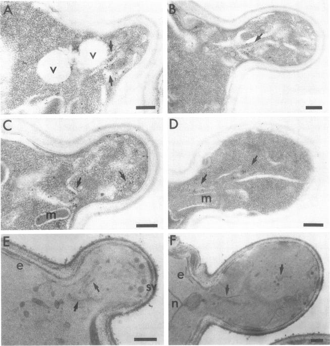
The membrane compartments responsible for Golgi functions in wild-type Saccharomyces cerevisiae were identified and characterized by immunoelectron microscopy. Using improved fixation methods, Golgi compartments were identified by labeling with antibodies specific for alpha 1-6 mannose linkages, the Sec7 protein, or the Ypt1 protein. The compartments labeled by each of these antibodies appear as disk-like structures that are apparently surrounded by small vesicles. Yeast Golgi typically are seen as single, isolated cisternae, generally not arranged into parallel stacks. The location of the Golgi structures was monitored by immunoelectron microscopy through the yeast cell cycle. Several Golgi compartments, apparently randomly distributed, were always observed in mother cells. During the initiation of new daughter cells, additional Golgi structures cluster just below the site of bud emergence. These Golgi enter daughter cells at an early stage, raising the possibility that much of the bud's growth might be due to secretory vesicles formed as well as consumed entirely within the daughter. During cytokinesis, the Golgi compartments are concentrated near the site of cell wall synthesis. Clustering of Golgi both at the site of bud formation and at the cell septum suggests that these organelles might be directed toward sites of rapid cell surface growth.
通过免疫电子显微镜鉴定并表征了野生型酿酒酵母中负责高尔基体功能的膜区室。采用改进的固定方法,通过用对α1-6甘露糖连接、Sec7蛋白或Ypt1蛋白特异的抗体进行标记来鉴定高尔基体区室。被这些抗体各自标记的区室呈现为盘状结构,显然被小泡所包围。酵母高尔基体通常被视为单个、孤立的扁平囊,一般不排列成平行堆叠。通过免疫电子显微镜在酵母细胞周期中监测高尔基体结构的位置。在母细胞中总是观察到几个高尔基体区室,明显呈随机分布。在新子细胞起始期间,额外的高尔基体结构聚集在芽出现位点的正下方。这些高尔基体在早期进入子细胞,这增加了一种可能性,即芽的大部分生长可能归因于在子细胞内形成并完全消耗的分泌小泡。在胞质分裂期间,高尔基体区室集中在细胞壁合成位点附近。高尔基体在芽形成位点和细胞隔膜处的聚集表明,这些细胞器可能被导向细胞表面快速生长的位点。