LISTGARTEN M A, LOESCHE W J, SOCRANSKY S S
J Bacteriol. 1963 Apr;85(4):932-9. doi: 10.1128/jb.85.4.932-939.1963.
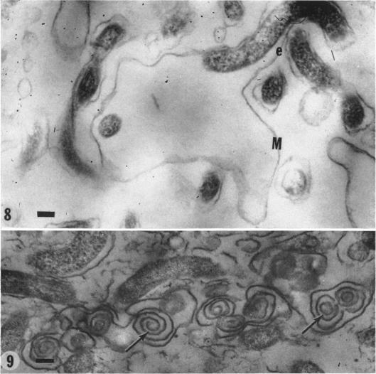
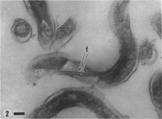
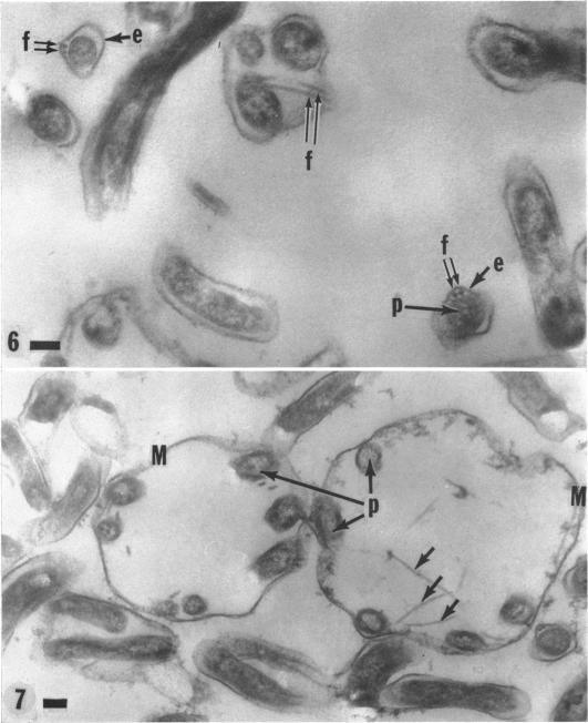
Listgarten, M. A. (Harvard School of Dental Medicine and Forsyth Dental Infirmary, Boston, Mass.), W. J. Loesche, and S. S. Socransky. Morphology of Treponema microdentium as revealed by electron microscopy of ultrathin sections. J. Bacteriol. 85:932-939. 1963.-Broth cultures of a strain of Treponema microdentium were harvested on Millipore filters, fixed in osmic acid, and sectioned for electron microscopy. The sections revealed that the spirochetes had an axial filament, made up of two fibrils approximately 150 A in diameter, which was situated between an external envelope approximately 140 A in thickness and a protoplasmic cylinder. The protoplasmic cylinder had a cross-sectional diameter of 100 to 200 mmu, and was surrounded by a double "membrane" consisting of two 40-A electron-dense structures separated by a 45-A space. Cross-sections of spirochetal "granules" revealed that the limiting membrane was continuous with the outer envelope of the spirochetes, and surrounded the protoplasmic cylinder and axial filament.
利斯特加滕,M. A.(马萨诸塞州波士顿哈佛牙医学院和福赛斯牙科医院),W. J. 洛舍,以及S. S. 索克兰斯基。超薄切片电子显微镜揭示的微小密螺旋体形态。《细菌学杂志》85:932 - 939。1963年。——将微小密螺旋体菌株的肉汤培养物收集在微孔滤膜上,用锇酸固定,然后切片用于电子显微镜观察。切片显示,螺旋体有一条轴丝,由两条直径约150埃的原纤维组成,位于厚度约140埃的外膜和原生质圆柱体之间。原生质圆柱体的横截面直径为100至200毫微米,被由两个40埃电子致密结构组成、中间间隔45埃空间的双层“膜”所包围。螺旋体“颗粒”的横截面显示,限制膜与螺旋体的外膜连续,并包围着原生质圆柱体和轴丝。