LISTGARTEN M A, SOCRANSKY S S
J Bacteriol. 1964 Oct;88(4):1087-103. doi: 10.1128/jb.88.4.1087-1103.1964.
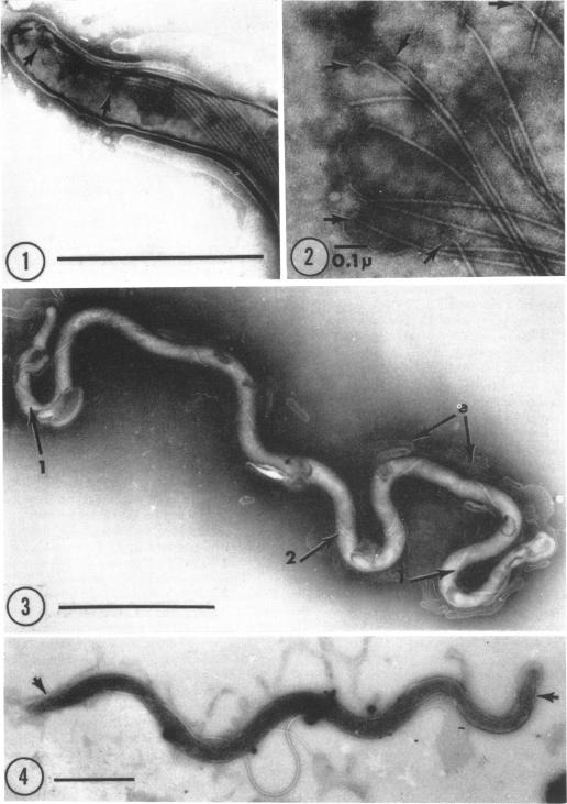
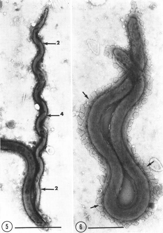
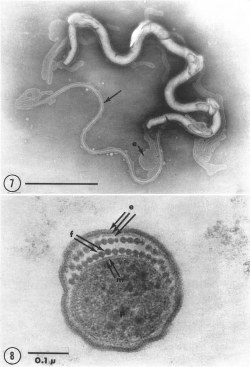
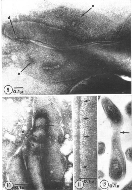
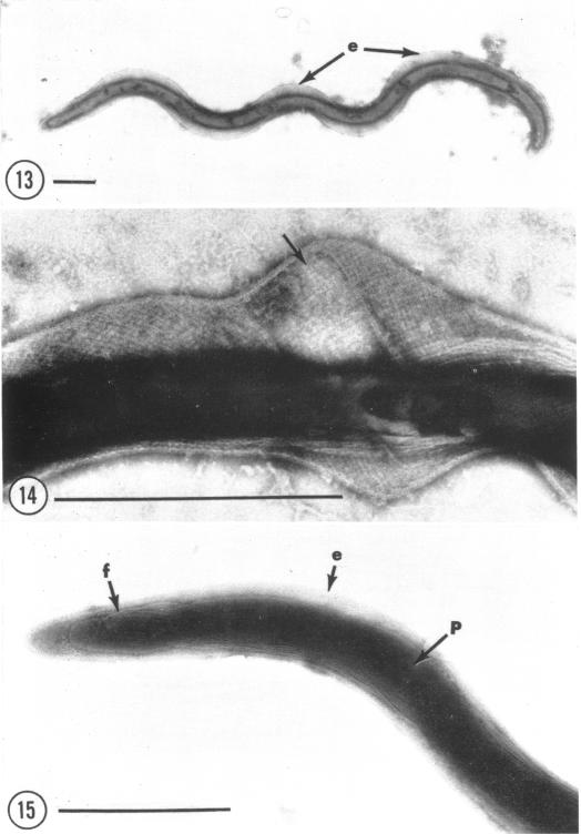
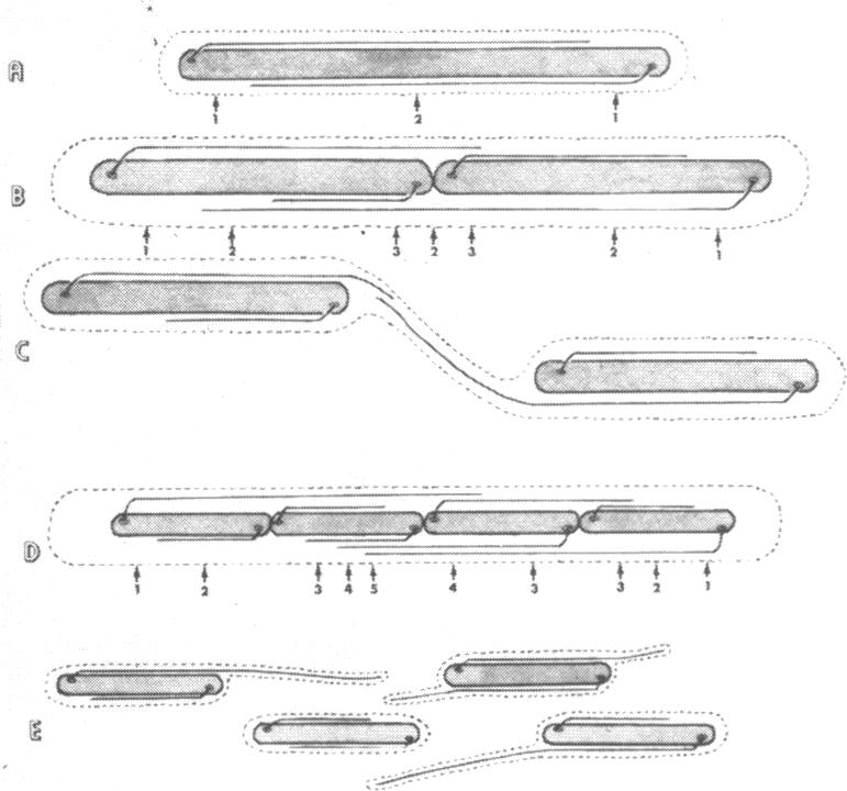
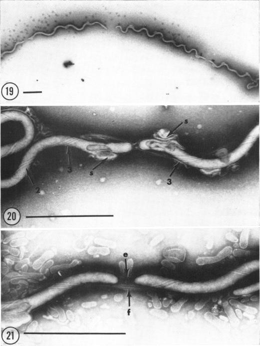
Listgarten, M. A. (Harvard School of Dental Medicine and Forsyth Dental Center, Boston, Mass.), and S. S. Socransky. Electron microscopy of axial fibrils, outer envelope, and cell division of certain oral spirochetes. J. Bacteriol. 88:1087-1103. 1964.-The ultrastructure of axial fibrils and outer envelopes of a number of oral spirochetes was studied in thin sections and by negative contrast. The axial fibrils measured 150 to 200 A in diameter. Only one end of each fibril was inserted subterminally into the protoplasmic cylinder by means of a 400 A wide disc. The free ends of fibrils inserted near one end of the cylinder extended toward, and overlapped in close apposition, the free ends of fibrils inserted at the other end. In thin sections, some axial fibrils showed a substructure, suggestive of a dense central core. The outer envelopes of most spirochetes appeared to consist of 80 A wide polygonal structural subunits. However, in one large spirochete, the outer envelope demonstrated a "pin-striped" pattern. Cell division in a pure culture of Treponema microdentium was studied by negative contrast. Results suggested that this organism divides by transverse fission, the outer envelope being last to divide. During the course of division, new axial fibrils appeared to originate on either side of the point of constriction of the protoplasmic cylinder. Flagellalike extensions which were found in rapidly dividing organisms were due to protruding axial fibrils, and appeared to be the result of cell division. Some evidence is presented to support the concept of a homologous origin for axial fibrils and flagella.
利斯特加滕,M. A.(哈佛牙医学院和福赛思牙科中心,马萨诸塞州波士顿),以及S. S. 索克兰斯基。某些口腔螺旋体的轴丝、外膜及细胞分裂的电子显微镜观察。《细菌学杂志》88:1087 - 1103。1964年。——通过超薄切片和负染色法研究了多种口腔螺旋体的轴丝和外膜的超微结构。轴丝直径为150至200埃。每根轴丝只有一端通过一个400埃宽的盘状结构亚末端插入原生质圆柱体。插入圆柱体一端附近的轴丝自由端朝着插入另一端的轴丝自由端延伸,并紧密并列重叠。在超薄切片中,一些轴丝显示出亚结构,提示有一个致密的中央核心。大多数螺旋体的外膜似乎由80埃宽的多边形结构亚基组成。然而,在一种大型螺旋体中,外膜呈现出“细条纹”图案。通过负染色法研究了微小密螺旋体纯培养物中的细胞分裂。结果表明,这种生物体通过横向分裂进行繁殖,外膜最后分裂。在分裂过程中,新的轴丝似乎起源于原生质圆柱体缢缩点的两侧。在快速分裂的生物体中发现的鞭毛样延伸是由于轴丝突出所致,似乎是细胞分裂的结果。提供了一些证据来支持轴丝和鞭毛同源起源的概念。