• 文献检索
  • 文档翻译
  • 深度研究
  • 学术资讯
  • Suppr Zotero 插件Zotero 插件
  • 邀请有礼
  • 套餐&价格
  • 历史记录
应用&插件
Suppr Zotero 插件Zotero 插件浏览器插件Mac 客户端Windows 客户端微信小程序
定价
高级版会员购买积分包购买API积分包
服务
文献检索文档翻译深度研究API 文档MCP 服务
关于我们
关于 Suppr公司介绍联系我们用户协议隐私条款
关注我们

Suppr 超能文献

核心技术专利:CN118964589B侵权必究
粤ICP备2023148730 号-1Suppr @ 2026

文献检索

告别复杂PubMed语法,用中文像聊天一样搜索,搜遍4000万医学文献。AI智能推荐,让科研检索更轻松。

立即免费搜索

文件翻译

保留排版,准确专业,支持PDF/Word/PPT等文件格式,支持 12+语言互译。

免费翻译文档

深度研究

AI帮你快速写综述,25分钟生成高质量综述,智能提取关键信息,辅助科研写作。

立即免费体验

密螺旋体的超微结构研究:梅毒螺旋体(Nichols株)、齿垢密螺旋体和赖氏密螺旋体的轴丝位置及一些尺寸

Ultrastructural studies of treponemes: location of axial filaments and some dimensions of Treponema pallidum (Nichols strain), Treponema denticola, and Treponema reiteri.

作者信息

Sykes J A, Miller J N

出版信息

Infect Immun. 1973 Jan;7(1):100-10. doi: 10.1128/iai.7.1.100-110.1973.

DOI:10.1128/iai.7.1.100-110.1973
PMID:4570277
原文链接:https://pmc.ncbi.nlm.nih.gov/articles/PMC422644/
Abstract

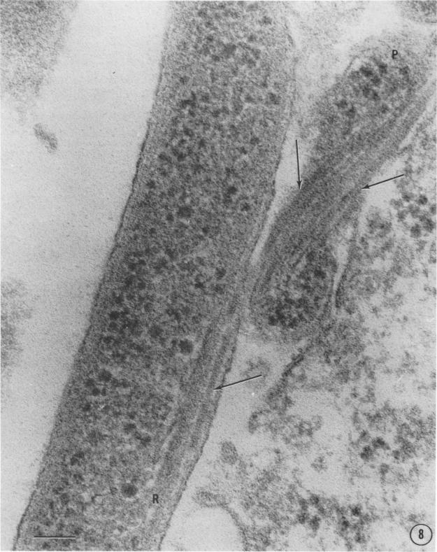
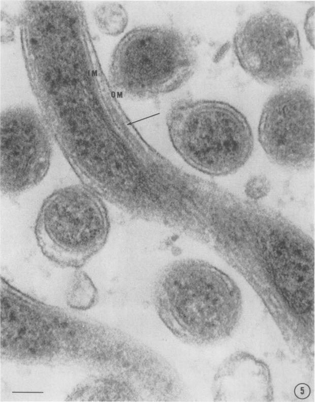
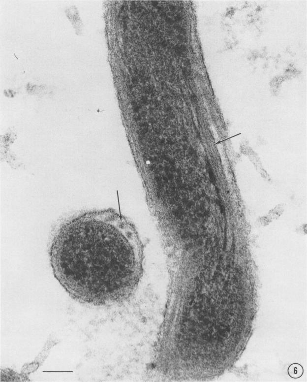
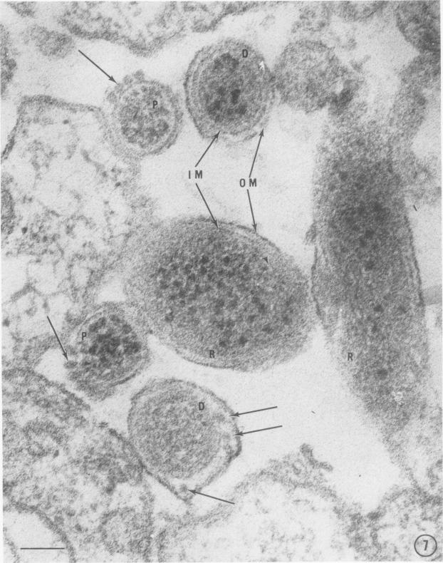
Ultrathin sections of Treponema pallidum (Nichols strain), T. denticola (microdentium), and T. reiteri have been studied in the electron microscope to determine the location of the axial filaments and some of the dimensions of these organisms. The axial filaments of T. pallidum (Nichols strain) have been seen to be tubular in cross section with an overall diameter of 21.0 +/- 0.73 nm, and an electron-lucent core of 8.0 nm. The filaments were found to lie on the outside of the organism which had only one membranous structure surrounding the protoplasmic core. These findings were in contrast to those obtained for T. denticola and T. reiteri where the axial filaments did not exhibit a hollow core and were located between an outer membrane and an inner membrane surrounding the protoplasmic core. The outside diameter of T. denticola was determined to be 224.9 +/- 2.83 nm, and that of T. reiteri as 331.0 +/- 4.15 nm, contrasting with T. pallidum (Nichols strain) which had a diameter of 163.0 +/- 1.9 nm.

摘要

对梅毒螺旋体(Nichols株)、齿垢密螺旋体和Reiter密螺旋体的超薄切片进行了电子显微镜研究,以确定轴丝的位置以及这些微生物的一些尺寸。已观察到梅毒螺旋体(Nichols株)的轴丝在横切面上呈管状,总直径为21.0±0.73纳米,有一个8.0纳米的电子透明核心。发现这些丝位于该微生物的外部,该微生物只有一层围绕原生质核心的膜结构。这些发现与齿垢密螺旋体和Reiter密螺旋体的情况形成对比,在后者中,轴丝没有中空核心,位于围绕原生质核心的外膜和内膜之间。齿垢密螺旋体的外径测定为224.9±2.83纳米,Reiter密螺旋体的外径为331.0±-4.15纳米,与直径为163.0±1.9纳米的梅毒螺旋体(Nichols株)形成对比。

https://cdn.ncbi.nlm.nih.gov/pmc/blobs/8f28/422644/cf72887fa65a/iai00253-0119-a.jpg
https://cdn.ncbi.nlm.nih.gov/pmc/blobs/8f28/422644/02301bf32d23/iai00253-0113-a.jpg
https://cdn.ncbi.nlm.nih.gov/pmc/blobs/8f28/422644/fa65be4c8a08/iai00253-0113-b.jpg
https://cdn.ncbi.nlm.nih.gov/pmc/blobs/8f28/422644/ade25ea366cd/iai00253-0115-a.jpg
https://cdn.ncbi.nlm.nih.gov/pmc/blobs/8f28/422644/4985d726ea46/iai00253-0115-b.jpg
https://cdn.ncbi.nlm.nih.gov/pmc/blobs/8f28/422644/6d156bf180dc/iai00253-0116-a.jpg
https://cdn.ncbi.nlm.nih.gov/pmc/blobs/8f28/422644/5e31be494235/iai00253-0117-a.jpg
https://cdn.ncbi.nlm.nih.gov/pmc/blobs/8f28/422644/39fcc95be98d/iai00253-0118-a.jpg
https://cdn.ncbi.nlm.nih.gov/pmc/blobs/8f28/422644/cf72887fa65a/iai00253-0119-a.jpg

相似文献

1
Ultrastructural studies of treponemes: location of axial filaments and some dimensions of Treponema pallidum (Nichols strain), Treponema denticola, and Treponema reiteri.密螺旋体的超微结构研究:梅毒螺旋体(Nichols株)、齿垢密螺旋体和赖氏密螺旋体的轴丝位置及一些尺寸
Infect Immun. 1973 Jan;7(1):100-10. doi: 10.1128/iai.7.1.100-110.1973.
2
Helical conformation of Treponema pallidum (Nichols strain), Treponema paraluis-cuniculi, Treponema denticola, Borrelia turicatae, and unidentified oral spirochetes.梅毒螺旋体(Nichols株)、兔梅毒螺旋体、齿垢密螺旋体、杜氏疏螺旋体及未鉴定的口腔螺旋体的螺旋结构。
Infect Immun. 1981 May;32(2):937-40. doi: 10.1128/iai.32.2.937-940.1981.
3
Outer envelope of virulent Treponema pallidum.恶性梅毒螺旋体的外膜。
Infect Immun. 1973 Aug;8(2):291-5. doi: 10.1128/iai.8.2.291-295.1973.
4
Treponema pallidum (Nichols strain) in tissue cultures: cellular attachment, entry, and survival.梅毒螺旋体(Nichols株)在组织培养中的细胞附着、进入及存活情况
Infect Immun. 1975 May;11(5):1133-40. doi: 10.1128/iai.11.5.1133-1140.1975.
5
Intracellular location of Treponema pallidum (Nichols strain) in the rabbit testis.梅毒螺旋体(Nichols株)在兔睾丸中的细胞内定位。
Infect Immun. 1971 Sep;4(3):307-14. doi: 10.1128/iai.4.3.307-314.1971.
6
Ultrastructure of Treponema pallidum Nichols following lysis by physical and chemical methods. II. Axial filaments.
Arch Mikrobiol. 1971;76(4):325-40. doi: 10.1007/BF00408529.
7
Immunity in experimental syphilis. IV. Serological reactivity of antigens extracted from gamm-irradiated Treponema pallidum and Treponema reiteri.实验性梅毒中的免疫。IV. 从经γ射线照射的梅毒螺旋体和里氏螺旋体中提取的抗原的血清学反应性。
J Bacteriol. 1966 Feb;91(2):583-7. doi: 10.1128/jb.91.2.583-587.1966.
8
Electron microscopy of treponema pallidum Nichols.梅毒螺旋体Nichols株的电子显微镜检查。
Acta Pathol Microbiol Scand. 1968;74(2):241-58. doi: 10.1111/j.1699-0463.1968.tb03477.x.
9
The ultrastructure of treponema pallidum isolated from human chancres. Morphologic variations from Nichols' strain.从人类溃疡中分离出的梅毒螺旋体的超微结构。与Nichols菌株的形态学差异。
Acta Derm Venereol. 1985;65(5):367-73.
10
Light and electron microscopy of rabbit testes infected with Treponema pallidum (Nichols strain): nature of deposited mucopolysaccharides and localisation of treponemes.感染梅毒螺旋体(Nichols株)的兔睾丸的光镜和电镜观察:沉积的粘多糖的性质及梅毒螺旋体的定位
Genitourin Med. 1987 Oct;63(5):297-304. doi: 10.1136/sti.63.5.297.

引用本文的文献

1
Biological basis for syphilis.梅毒的生物学基础。
Clin Microbiol Rev. 2006 Jan;19(1):29-49. doi: 10.1128/CMR.19.1.29-49.2006.
2
Outer envelope of virulent Treponema pallidum.恶性梅毒螺旋体的外膜。
Infect Immun. 1973 Aug;8(2):291-5. doi: 10.1128/iai.8.2.291-295.1973.
3
Unusual fibrils from the spirochete-like sex ratio organism.来自类螺旋体性别比例生物的异常纤丝。

本文引用的文献

1
ELECTRON MICROSCOPY OF AXIAL FIBRILS, OUTER ENVELOPE, AND CELL DIVISION OF CERTAIN ORAL SPIROCHETES.某些口腔螺旋体的轴丝、外膜及细胞分裂的电子显微镜观察
J Bacteriol. 1964 Oct;88(4):1087-103. doi: 10.1128/jb.88.4.1087-1103.1964.
2
MORPHOLOGY OF TREPONEMA MICRODENTIUM AS REVEALED BY ELECTRON MICROSCOPY OF ULTRATHIN SECTIONS.超薄切片电子显微镜观察微小密螺旋体的形态学
J Bacteriol. 1963 Apr;85(4):932-9. doi: 10.1128/jb.85.4.932-939.1963.
3
[Electron microscope study of the external and internal structure of the Reiter treponema].
J Bacteriol. 1974 Feb;117(2):904-6. doi: 10.1128/jb.117.2.904-906.1974.
4
Treponema phagedenis has at least two proteins residing together on its periplasmic flagella.溃蚀性密螺旋体在其周质鞭毛上至少有两种蛋白质共存。
J Bacteriol. 1986 Apr;166(1):105-12. doi: 10.1128/jb.166.1.105-112.1986.
5
The surface of virulent Treponema pallidum: resistance to antibody binding in the absence of complement and surface association of recombinant antigen 4D.毒力梅毒螺旋体的表面:在无补体情况下对抗体结合的抗性及重组抗原4D的表面结合
Infect Immun. 1986 May;52(2):579-85. doi: 10.1128/iai.52.2.579-585.1986.
6
Role of disulfide bonds in the oligomeric structure and protease resistance of recombinant and native Treponema pallidum surface antigen 4D.二硫键在重组和天然梅毒螺旋体表面抗原4D的寡聚结构及蛋白酶抗性中的作用
J Bacteriol. 1987 Apr;169(4):1365-71. doi: 10.1128/jb.169.4.1365-1371.1987.
7
Changes in the cell surface properties of Treponema pallidum that occur during in vitro incubation of freshly extracted organisms.新鲜提取的梅毒螺旋体在体外培养过程中细胞表面特性的变化。
Infect Immun. 1987 Sep;55(9):2255-61. doi: 10.1128/iai.55.9.2255-2261.1987.
8
Interactions of Treponema pallidum with endothelial cell monolayers.梅毒螺旋体与内皮细胞单层的相互作用。
Eur J Epidemiol. 1989 Mar;5(1):15-21. doi: 10.1007/BF00145039.
9
Antigenic interrelationship between endoflagella of Treponema phagedenis biotype Reiter and Treponema pallidum (Nichols): molecular characterization of endoflagellar proteins.噬菌性密螺旋体生物变种赖特株与梅毒螺旋体(Nichols株)内鞭毛之间的抗原相互关系:内鞭毛蛋白的分子特征
Infect Immun. 1986 Dec;54(3):626-34. doi: 10.1128/iai.54.3.626-634.1986.
10
The bent-end morphology of Treponema phagedenis is associated with short, left-handed, periplasmic flagella.溃蚀性密螺旋体的弯曲末端形态与短的、左旋的周质鞭毛有关。
J Bacteriol. 1991 Aug;173(15):4820-6. doi: 10.1128/jb.173.15.4820-4826.1991.
[赖特疏螺旋体外在与内在结构的电子显微镜研究]
Ann Inst Pasteur (Paris). 1963 Apr;104:496-501.
4
Electron microscopic studies of the morphology of pathogenic spirochaetes.致病性螺旋体形态的电子显微镜研究。
J Pathol Bacteriol. 1955 Jan-Apr;69(1-2):117-28. doi: 10.1002/path.1700690117.
5
Intracellular location of Treponema pallidum (Nichols strain) in the rabbit testis.梅毒螺旋体(Nichols株)在兔睾丸中的细胞内定位。
Infect Immun. 1971 Sep;4(3):307-14. doi: 10.1128/iai.4.3.307-314.1971.
6
Current concepts of the morphology and biology of Treponema pallidum based on electron microscopy.基于电子显微镜的梅毒螺旋体形态学与生物学的当前概念。
Br J Vener Dis. 1971 Oct;47(5):315-28. doi: 10.1136/sti.47.5.315.
7
An electron microscopic study of a syphilitic chancre. Engulfment of Treponema pallidum by plasma cells.梅毒初疮的电子显微镜研究。浆细胞对梅毒螺旋体的吞噬作用。
Arch Pathol. 1970 Aug;90(2):143-50.
8
Further studies of the morphology of Treponema pallidum under the electron microscope.梅毒螺旋体在电子显微镜下形态的进一步研究。
Br J Vener Dis. 1969 Jun;45(2):87-116. doi: 10.1136/sti.45.2.87.
9
Electron microscopic observations on the lesions of condyloma latum.扁平湿疣病变的电子显微镜观察
Br J Dermatol. 1969 May;81(5):367-74. doi: 10.1111/j.1365-2133.1969.tb14000.x.
10
Electron microscopy of Treponema pallidum occurring in a human primary lesion.发生于人类原发性损害中的梅毒螺旋体的电子显微镜检查。
J Bacteriol. 1969 Feb;97(2):951-5. doi: 10.1128/jb.97.2.951-955.1969.