Takeda A, Jimi T, Wakayama Y, Misugi N, Miyake S, Kumagai T
Department of Clinical Pathology, Showa University Fujigaoka Hospital, Showa University School of Medicine, Yokohama, Japan.
Biochem J. 1992 Dec 1;288 ( Pt 2)(Pt 2):643-8. doi: 10.1042/bj2880643.
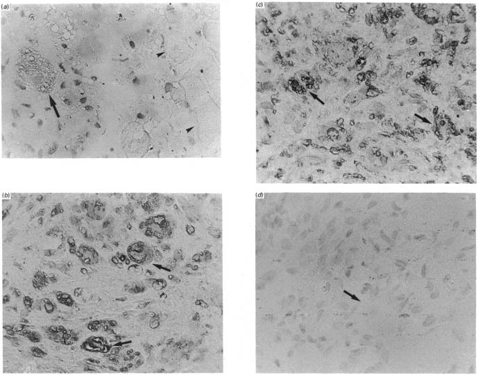
The activities and contents of the lysosomal cysteine proteinases cathepsins B, H and L were examined in xenografts of biopsied muscles transplanted from age-matched normal subjects and Duchenne-muscular-dystrophy (DMD) patients into nude mice. The activity of cathepsin B increased 9-fold and that of B-plus-L increased 24-fold in the first week after transplantation in normal muscle xenografts. By the third week, the activity of cathepsin B increased a total of 20-fold and B-plus-L increased to 36-fold the original level. The activity levels of cathepsin B, B-plus-L, H and D, and acid phosphatase in normal and DMD xenografts were not significantly different when compared 2 weeks after transplantation. However, the protein content of cathepsin B in DMD muscle xenografts was more than 3-fold that of normal xenografts at 2 weeks. The profile of cathepsin H activity in normal muscle xenografts was different than those of cathepsins B and B-plus-L. In the first week, the cathepsin H diminished sharply to about one-third of the biopsied muscle level and then, by 3 weeks after transplantation, it had increased slightly to about half the original level. The amount of endogenous cysteine-proteinase inhibitor changed in parallel with the activity of cathepsins B and B-plus-L. Cathepsins B and H, but not cathepsin L, were found immunohistochemically in regenerating muscle fibres of normal and DMD xenografts 2 weeks after transplantation. Staining of cathepsin B in DMD xenografts was slightly stronger than that in normal subjects. There was no immunostaining in degenerating or necrotic muscle fibres 2 weeks after transplantation. Western-blot analysis revealed that the cathepsin B band at 29 kDa was increased in normal xenografts 2 and 3 weeks after transplantation. Also, 2 weeks after transplantation the staining intensity of this band was slightly stronger in DMD xenografts than in normal xenografts. These results suggest that cathepsin B participates in the regeneration of transplanted muscle, both normal and DMD, and in the DMD muscle fibre-wasting processes, during regeneration.
在将年龄匹配的正常受试者和杜兴氏肌营养不良症(DMD)患者的活检肌肉移植到裸鼠体内形成的异种移植中,对溶酶体半胱氨酸蛋白酶组织蛋白酶B、H和L的活性及含量进行了检测。在正常肌肉异种移植中,移植后第一周组织蛋白酶B的活性增加了9倍,B加L的活性增加了24倍。到第三周时,组织蛋白酶B的活性总共增加了20倍,B加L增加到原来水平的36倍。移植2周后比较时,正常和DMD异种移植中组织蛋白酶B、B加L、H和D以及酸性磷酸酶的活性水平没有显著差异。然而,在2周时,DMD肌肉异种移植中组织蛋白酶B的蛋白质含量是正常异种移植的3倍多。正常肌肉异种移植中组织蛋白酶H的活性谱与组织蛋白酶B和B加L的不同。在第一周,组织蛋白酶H急剧下降至活检肌肉水平的约三分之一,然后在移植后3周时,它略有增加至原来水平的约一半。内源性半胱氨酸蛋白酶抑制剂的量与组织蛋白酶B和B加L的活性平行变化。移植2周后,在正常和DMD异种移植的再生肌纤维中通过免疫组织化学方法检测到了组织蛋白酶B和H,但未检测到组织蛋白酶L。DMD异种移植中组织蛋白酶B的染色略强于正常受试者。移植2周后,在退化或坏死的肌纤维中没有免疫染色。蛋白质印迹分析显示,移植后2周和3周,正常异种移植中29 kDa的组织蛋白酶B条带增加。同样,移植2周后,DMD异种移植中该条带的染色强度略强于正常异种移植。这些结果表明,组织蛋白酶B参与了正常和DMD移植肌肉的再生,以及DMD肌肉纤维在再生过程中的消耗过程。