Karbowski Mariusz, Arnoult Damien, Chen Hsiuchen, Chan David C, Smith Carolyn L, Youle Richard J
Biochemistry Section, Surgical Neurology Branch, National Institute of Neurological Disorders and Stroke, National Institutes of Health, Bethesda, MD 20892, USA.
J Cell Biol. 2004 Feb 16;164(4):493-9. doi: 10.1083/jcb.200309082. Epub 2004 Feb 9.
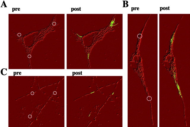
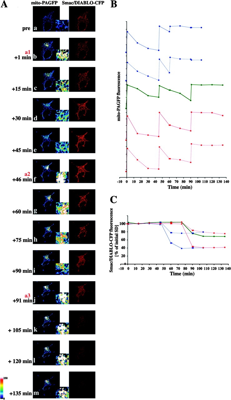
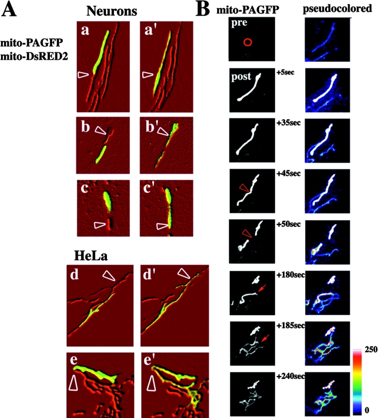
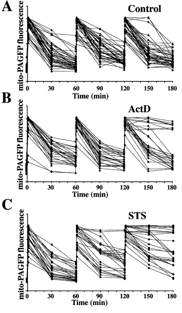
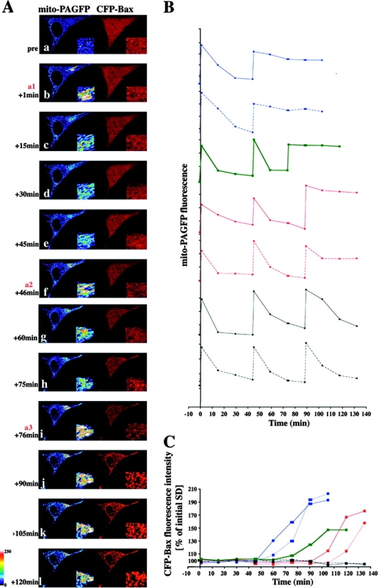
A dynamic balance of organelle fusion and fission regulates mitochondrial morphology. During apoptosis this balance is altered, leading to an extensive fragmentation of the mitochondria. Here, we describe a novel assay of mitochondrial dynamics based on confocal imaging of cells expressing a mitochondrial matrix-targeted photoactivable green fluorescent protein that enables detection and quantification of organelle fusion in living cells. Using this assay, we visualize and quantitate mitochondrial fusion rates in healthy and apoptotic cells. During apoptosis, mitochondrial fusion is blocked independently of caspase activation. The block in mitochondrial fusion occurs within the same time range as Bax coalescence on the mitochondria and outer mitochondrial membrane permeabilization, and it may be a consequence of Bax/Bak activation during apoptosis.
细胞器融合与分裂的动态平衡调节线粒体形态。在细胞凋亡过程中,这种平衡会发生改变,导致线粒体广泛碎片化。在此,我们描述了一种基于共聚焦成像的线粒体动力学新检测方法,该方法用于对表达线粒体基质靶向的光激活绿色荧光蛋白的细胞进行成像,能够在活细胞中检测和定量细胞器融合。利用该检测方法,我们可视化并定量了健康细胞和凋亡细胞中的线粒体融合速率。在细胞凋亡过程中,线粒体融合被阻断,且与半胱天冬酶激活无关。线粒体融合的阻断发生在与 Bax 在线粒体上聚集以及线粒体外膜通透性改变相同的时间范围内,它可能是细胞凋亡过程中 Bax/Bak 激活的结果。