Yang G C, Nieto R, Stachura I, Gallo G R
Department of Pathology, New York University Medical Center, New York 10016.
Am J Pathol. 1992 Aug;141(2):409-19.
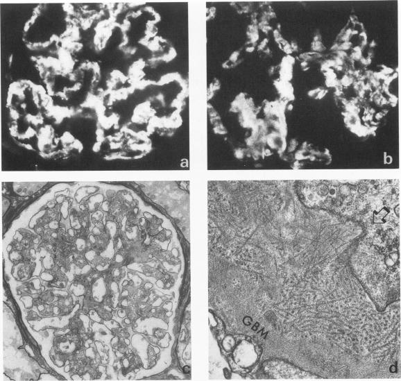
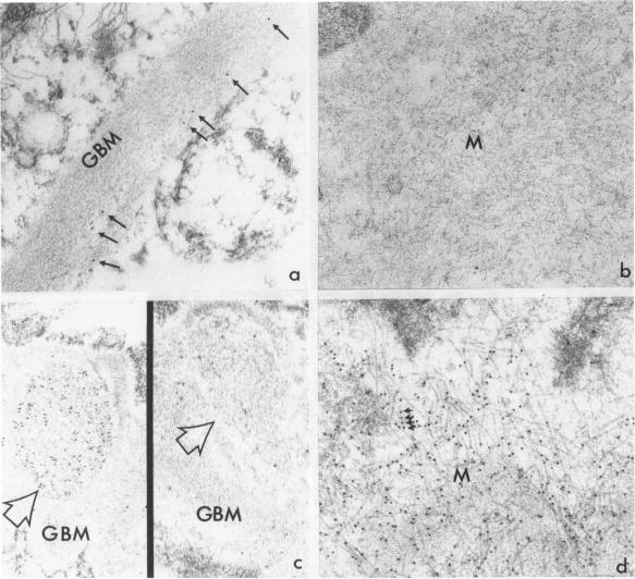
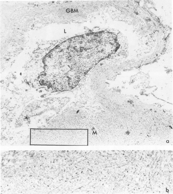
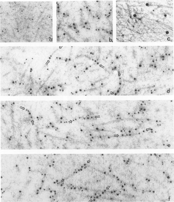
Renal biopsies from seven patients with Congo red-negative amyloid-like fibrillary glomerulopathy (FGP) were examined by protein A gold immuno-electron microscopy. Ultrastructurally, the fibrils in all cases exhibited positive immunostaining for IgG, both Ig light chains, C3, and amyloid P component (AP), but did not show positive immunostaining for glomerular basement membrane (GBM)-associated proteins (collagen type IV and heparan-sulfate proteoglycans) or microfibril-associated proteins (fibronectin and fibrillin). In a triple-label study, AP and IgG were colocalized along the same fibril, whereas the gold probes for the detection of collagen type IV were absent. The results suggest that the fibrils are comprised of polyclonal IgG and C3 that bind AP. AP was immunolocalized sparsely but regularly along the lamina rara interna of normal GBM. AP was absent in the fibrils in a case of diabetic glomerulopathy, was scattered randomly without specificity for the electron-dense deposits in the GBM of membranous glomerulopathy, and lined up regularly along the fibrils in amyloid deposits. FGP is an entity in which the fibrils bind AP but lack the beta-pleated sheet structure necessary for Congo red staining that is typical of amyloid.
对7例刚果红阴性淀粉样纤维性肾小球病(FGP)患者的肾活检组织进行了蛋白A金免疫电子显微镜检查。超微结构显示,所有病例中的纤维对IgG、两种Ig轻链、C3和淀粉样P成分(AP)均呈阳性免疫染色,但对肾小球基底膜(GBM)相关蛋白(IV型胶原和硫酸乙酰肝素蛋白聚糖)或微纤维相关蛋白(纤连蛋白和原纤蛋白)未呈阳性免疫染色。在一项三重标记研究中,AP和IgG沿同一纤维共定位,而用于检测IV型胶原的金探针未出现。结果表明,这些纤维由结合AP的多克隆IgG和C3组成。AP在正常GBM的内疏松层呈稀疏但规则的免疫定位。在糖尿病性肾小球病病例中,纤维中不存在AP;在膜性肾小球病的GBM电子致密沉积物中,AP随机分布且无特异性;在淀粉样沉积物的纤维中,AP沿纤维规则排列。FGP是一种纤维结合AP但缺乏刚果红染色所需的典型淀粉样β折叠结构的疾病实体。