• 文献检索
  • 文档翻译
  • 深度研究
  • 学术资讯
  • Suppr Zotero 插件Zotero 插件
  • 邀请有礼
  • 套餐&价格
  • 历史记录
应用&插件
Suppr Zotero 插件Zotero 插件浏览器插件Mac 客户端Windows 客户端微信小程序
定价
高级版会员购买积分包购买API积分包
服务
文献检索文档翻译深度研究API 文档MCP 服务
关于我们
关于 Suppr公司介绍联系我们用户协议隐私条款
关注我们

Suppr 超能文献

核心技术专利:CN118964589B侵权必究
粤ICP备2023148730 号-1Suppr @ 2026

文献检索

告别复杂PubMed语法,用中文像聊天一样搜索,搜遍4000万医学文献。AI智能推荐,让科研检索更轻松。

立即免费搜索

文件翻译

保留排版,准确专业,支持PDF/Word/PPT等文件格式,支持 12+语言互译。

免费翻译文档

深度研究

AI帮你快速写综述,25分钟生成高质量综述,智能提取关键信息,辅助科研写作。

立即免费体验

纤维性肾沉积物与肾炎。

Fibrillary renal deposits and nephritis.

作者信息

Duffy J L, Khurana E, Susin M, Gomez-Leon G, Churg J

出版信息

Am J Pathol. 1983 Dec;113(3):279-90.

PMID:6359891
原文链接:https://pmc.ncbi.nlm.nih.gov/articles/PMC1916347/
Abstract

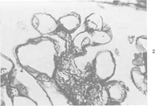
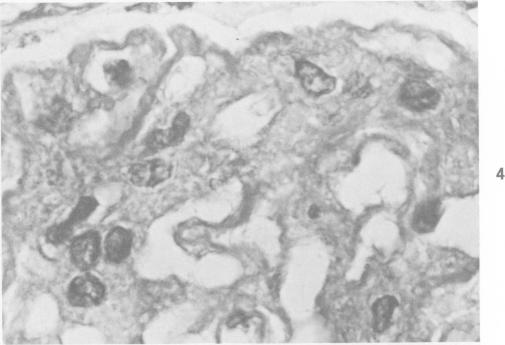
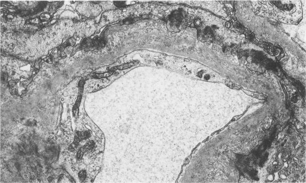
Fibrillary renal deposits and nephritis. The authors have studied 8 patients whose glomeruli contain abundant fibrils in their mesangial matrix and basement membranes. Although the location of these fibrils is very similar to that of amyloid, they are about twice the size of amyloid fibrils, averaging 20 nm in width, and fail to react as amyloid does with special stains. Immunofluorescence-microscopic studies are usually positive with antiserums to IgG, often IgM, and in some cases IgA, and also kappa and lambda light chains, C3, and C4. The fibrils are associated with diffuse mesangial widening and increased mesangial matrix strands. Although peripheral glomerular capillary walls appear to be spared initially, their eventual involvement leads to glomerular capillary collapse and glomerular obsolescence. Crescent formation occurred in 5 cases, focally in 3 and diffusely in 2. Tubular basement membrane involvement was seen in 1 case. These patients exhibit hematuria, and proteinuria, and often hypertension and renal insufficiency. Proteinuria was in the nephrotic range in 3 patients in whom involvement of glomerular capillary basement membranes was extensive. Unless electron microscopy is applied to renal biopsies, these cases may be considered to represent mesangiocapillary or rapidly progressive glomerulonephritis, or amyloidosis. The nature of these fibrils is as yet not determined. It is likely that they have been called "atypical amyloidosis" in the past.

摘要

纤维性肾沉积物与肾炎。作者研究了8例患者,其肾小球系膜基质和基底膜中含有丰富的纤维。尽管这些纤维的位置与淀粉样蛋白非常相似,但它们的大小约为淀粉样纤维的两倍,平均宽度为20纳米,并且不像淀粉样蛋白那样对特殊染色有反应。免疫荧光显微镜研究通常对抗IgG血清呈阳性,常为IgM,在某些情况下为IgA,以及κ和λ轻链、C3和C4。这些纤维与弥漫性系膜增宽和系膜基质条索增加有关。尽管肾小球外周毛细血管壁最初似乎未受累,但它们最终的受累会导致肾小球毛细血管塌陷和肾小球荒废。5例出现新月体形成,3例为局灶性,2例为弥漫性。1例可见肾小管基底膜受累。这些患者表现为血尿、蛋白尿,常伴有高血压和肾功能不全。3例肾小球毛细血管基底膜广泛受累的患者蛋白尿处于肾病范围。除非对肾活检进行电子显微镜检查,否则这些病例可能被认为代表系膜毛细血管性或快速进展性肾小球肾炎,或淀粉样变性。这些纤维的性质尚未确定。过去它们可能被称为“非典型淀粉样变性”。

https://cdn.ncbi.nlm.nih.gov/pmc/blobs/92ae/1916347/dc8c467d592c/amjpathol00189-0026-a.jpg
https://cdn.ncbi.nlm.nih.gov/pmc/blobs/92ae/1916347/aee1d0e23d90/amjpathol00189-0021-a.jpg
https://cdn.ncbi.nlm.nih.gov/pmc/blobs/92ae/1916347/b997d23ef0c8/amjpathol00189-0021-b.jpg
https://cdn.ncbi.nlm.nih.gov/pmc/blobs/92ae/1916347/48fc82f58572/amjpathol00189-0021-c.jpg
https://cdn.ncbi.nlm.nih.gov/pmc/blobs/92ae/1916347/be3fed2304a9/amjpathol00189-0021-d.jpg
https://cdn.ncbi.nlm.nih.gov/pmc/blobs/92ae/1916347/8609255cd4df/amjpathol00189-0021-e.jpg
https://cdn.ncbi.nlm.nih.gov/pmc/blobs/92ae/1916347/47d054f8e2fd/amjpathol00189-0021-f.jpg
https://cdn.ncbi.nlm.nih.gov/pmc/blobs/92ae/1916347/77aaa008f211/amjpathol00189-0022-a.jpg
https://cdn.ncbi.nlm.nih.gov/pmc/blobs/92ae/1916347/44766f70f3f7/amjpathol00189-0023-a.jpg
https://cdn.ncbi.nlm.nih.gov/pmc/blobs/92ae/1916347/8748d9f028d8/amjpathol00189-0024-a.jpg
https://cdn.ncbi.nlm.nih.gov/pmc/blobs/92ae/1916347/ee15c27ede18/amjpathol00189-0025-a.jpg
https://cdn.ncbi.nlm.nih.gov/pmc/blobs/92ae/1916347/dc8c467d592c/amjpathol00189-0026-a.jpg

相似文献

1
Fibrillary renal deposits and nephritis.纤维性肾沉积物与肾炎。
Am J Pathol. 1983 Dec;113(3):279-90.
2
Glomerulonephropathy with amyloid-stain-negative microfibrillar glomerular deposits.伴有淀粉样蛋白染色阴性微纤维肾小球沉积物的肾小球病
Nephron. 1988;48(1):33-9. doi: 10.1159/000184865.
3
Lupus nephritis: correlation between light, electron microscopic and immunofluorescent findings and renal function.狼疮性肾炎:光镜、电镜及免疫荧光检查结果与肾功能的相关性
Clin Nephrol. 1976 Aug;6(2):340-51.
4
Immunotactoid glomerulopathy.免疫触须样肾小球病
Medicine (Baltimore). 1985 Jul;64(4):228-43. doi: 10.1097/00005792-198507000-00004.
5
Fibrillary glomerulonephritis: a report of 2 cases with extensive glomerular and tubular deposits.纤维样肾小球肾炎:2例伴有广泛肾小球和肾小管沉积物的病例报告
Hum Pathol. 2001 Jun;32(6):660-3. doi: 10.1053/hupa.2001.25005.
6
[Observations on the ultrastructure of non-amyloidotic fibrillary glomerulopathy].[非淀粉样纤维性肾小球病的超微结构观察]
Zhonghua Bing Li Xue Za Zhi. 1995 Jun;24(3):146-8.
7
Concurrent feline immune-complex nephritis. Tubular antigen-positive and renal amyloidosis.并发猫免疫复合物性肾炎。肾小管抗原阳性及肾淀粉样变性。
Arch Pathol Lab Med. 1979 Aug;103(9):475-8.
8
Immunofluorescence characterization of light chains in human nephropathies.人类肾病中轻链的免疫荧光特征分析
Virchows Arch A Pathol Anat Histopathol. 1988;412(6):591-4. doi: 10.1007/BF00844295.
9
[Clinicopathologic features of membranous nephropathy coexisting with IgA nephropathy].膜性肾病合并IgA肾病的临床病理特征
Zhonghua Bing Li Xue Za Zhi. 2007 Mar;36(3):171-4.
10
Monoclonal immunoglobulin G1-kappa fibrillary glomerulonephritis.单克隆免疫球蛋白G1-κ轻链纤维性肾小球肾炎
Mod Pathol. 1998 Jan;11(1):103-9.

引用本文的文献

1
Fibrillary Glomerulonephritis: Clinicopathological Characteristics and Outcome-Case Series From a Multicentre Australasian Cohort.纤维性肾小球肾炎:来自澳大利亚多中心队列的临床病理特征及预后——病例系列研究
Nephrology (Carlton). 2025 Apr;30(4):e70022. doi: 10.1111/nep.70022.
2
Renal Manifestations of Chronic Hepatitis C: A Review.慢性丙型肝炎的肾脏表现:综述
J Clin Med. 2024 Sep 18;13(18):5536. doi: 10.3390/jcm13185536.
3
Fibrillary Glomerulonephritis and DnaJ Homolog Subfamily B Member 9 (DNAJB9).纤维状肾小球肾炎和 DNAJ 同源物亚家族 B 成员 9(DNAJB9)。

本文引用的文献

1
The Mesangium of the Renal Glomerulus: Electron Microscopic Studies of Pathologic Alterations.肾小肾小球系膜:病理改变的电子显微镜研究
Am J Pathol. 1963 Oct;43(4):555-78.
2
Glomerular permeability. I. Ferritin transfer across the normal glomerular capillary wall.肾小球通透性。I. 铁蛋白穿过正常肾小球毛细血管壁的转运。
J Exp Med. 1961 Jan 1;113(1):47-66. doi: 10.1084/jem.113.1.47.
3
Electron microscopic observations on a fibrous component in amyloid of diverse origins.对不同来源淀粉样蛋白中一种纤维成分的电子显微镜观察。
Kidney360. 2020 Jul 8;1(9):1002-1013. doi: 10.34067/KID.0002532020. eCollection 2020 Sep 24.
4
HCV-Associated Nephropathies in the Era of Direct Acting Antiviral Agents.直接抗病毒药物时代的丙型肝炎病毒相关肾病
Front Med (Lausanne). 2019 Feb 8;6:20. doi: 10.3389/fmed.2019.00020. eCollection 2019.
5
DNAJB9 Is a Specific Immunohistochemical Marker for Fibrillary Glomerulonephritis.DNAJB9是纤维性肾小球肾炎的一种特异性免疫组织化学标志物。
Kidney Int Rep. 2017 Aug 8;3(1):56-64. doi: 10.1016/j.ekir.2017.07.017. eCollection 2018 Jan.
6
Clinical Features and Outcomes of a Racially Diverse Population with Fibrillary Glomerulonephritis.不同种族的纤维性肾小球肾炎患者的临床特征及预后
Am J Nephrol. 2017;45(3):248-256. doi: 10.1159/000455390. Epub 2017 Feb 4.
7
[Lupus nephritis with crystal structures in glomerulopathy ].狼疮性肾炎伴肾小球病变中的晶体结构
Pathologe. 2003 Oct;24(6):433-8. doi: 10.1007/s00292-003-0658-1.
8
Immunotactoid glomerulopathy in a child with Down syndrome.一名唐氏综合征患儿的免疫触须样肾小球病。
Pediatr Nephrol. 1993 Feb;7(1):86-8. doi: 10.1007/BF00861584.
9
Nephrotic syndrome and primary tubular amyloidosis in a child: a case report.一名儿童的肾病综合征和原发性肾小管淀粉样变性:病例报告
Pediatr Nephrol. 1989 Oct;3(4):454-7. doi: 10.1007/BF00850227.
10
Immunotactoid glomerulopathy with fingerprint immune deposits. A variant of lupus nephritis?伴有指纹状免疫沉积物的免疫触须样肾小球病。狼疮性肾炎的一种变异型?
Virchows Arch A Pathol Anat Histopathol. 1989;415(2):181-6. doi: 10.1007/BF00784357.
Nature. 1959 Apr 25;183(4669):1202-3. doi: 10.1038/1831202a0.
4
The fine structure of the renal glomerulus of the mouse.小鼠肾小球的精细结构。
J Biophys Biochem Cytol. 1955 Nov 25;1(6):551-66. doi: 10.1083/jcb.1.6.551.
5
Membranolysis by the ninth component of human complement.人补体第九成分所致的膜溶解作用。
Biochem Biophys Res Commun. 1981 Jun 16;100(3):1409-14. doi: 10.1016/0006-291x(81)91981-1.
6
Renal disease in essential mixed cryoglobulinaemia. Long-term follow-up of 44 patients.原发性混合性冷球蛋白血症中的肾脏疾病。44例患者的长期随访
Q J Med. 1981;50(197):1-30.
7
The value of electron microscopy in diagnostic pathology. Case 2.电子显微镜在诊断病理学中的价值。病例2。
Ultrastruct Pathol. 1980 Jan-Mar;1(1):79-83. doi: 10.3109/01913128009141400.
8
Microfibrils in glomerulopathies of children: an ultrastructural study.儿童肾小球病中的微原纤维:一项超微结构研究。
J Pathol. 1982 Feb;136(2):123-31. doi: 10.1002/path.1711360206.
9
Formation of transmembrane tubules by spontaneous polymerization of the hydrophilic complement protein C9.亲水性补体蛋白C9通过自发聚合形成跨膜小管。
Nature. 1982 Aug 5;298(5874):534-8. doi: 10.1038/298534a0.
10
Diabetic glomerulonephropathy: histopathologic, immunofluorescent, and ultrastructural studies of 16 cases.糖尿病肾小球肾病:16例的组织病理学、免疫荧光及超微结构研究
Hum Pathol. 1982 May;13(5):470-8. doi: 10.1016/s0046-8177(82)80030-0.