Gruber Günther, Greiner Richard H, Hlushchuk Ruslan, Aebersold Daniel M, Altermatt Hans J, Berclaz Gilles, Djonov Valentin
Department of Radiation Oncology, University of Bern, Switzerland.
Breast Cancer Res. 2004;6(3):R191-8. doi: 10.1186/bcr775. Epub 2004 Mar 9.
Hypoxia-inducible factor 1 alpha (hif-1alpha) furnishes tumor cells with the means of adapting to stress parameters like tumor hypoxia and promotes critical steps in tumor progression and aggressiveness. We investigated the role of hif-1alpha expression in patients with node-positive breast cancer.
Tumor samples from 77 patients were available for immunohistochemistry. The impact of hif-1alpha immunoreactivity on survival endpoints was determined by univariate and multivariate analyses, and correlations to clinicopathological characteristics were determined by cross-tabulations.
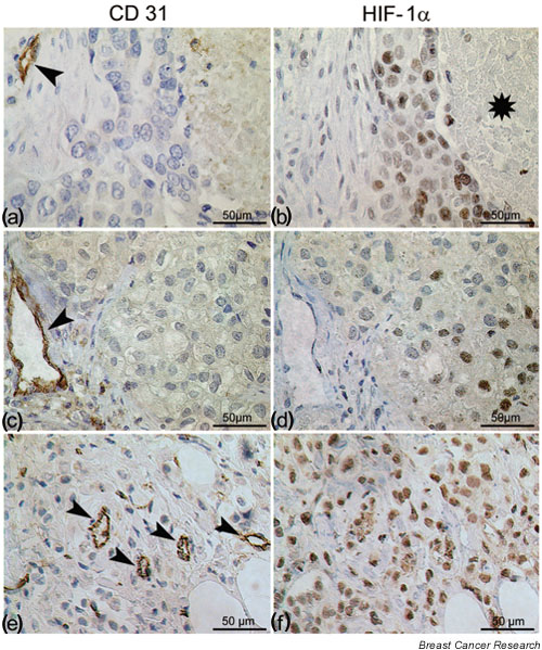
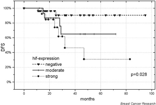
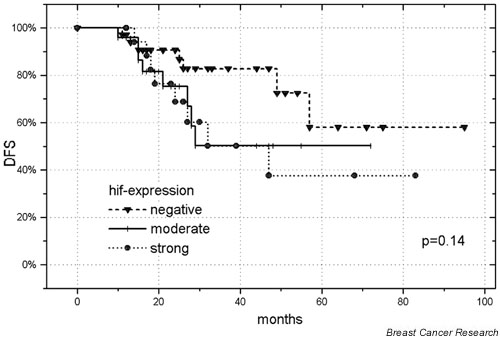
hif-1alpha was expressed in 56% (n = 43/77) of the patients. Its expression correlated with progesterone receptor negativity (P = 0.002). The Kaplan-Meier curves revealed significantly shorter distant metastasis-free survival (DMFS) (P = 0.04, log-rank) and disease-free survival (DFS) (P = 0.04, log-rank) in patients with increased hif-1alpha expression. The difference in overall survival (OS) did not attain statistical significance (5-year OS, 66% without hif-1alpha expression and 55% with hif-1alpha expression; P = 0.21). The multivariate analysis failed to reveal an independent prognostic value for hif-1alpha expression in the whole patient group. The only significant parameter for all endpoints was the T stage (T3/T4 versus T1/T2: DMFS, relative risk = 3.16, P = 0.01; DFS, relative risk = 2.57, P = 0.03; OS, relative risk = 3.03, P = 0.03). Restricting the univariate and multivariate analyses to T1/T2 tumors, hif-1alpha expression was a significant parameter for DFS and DMFS.
hif-1alpha is expressed in the majority of patients with node-positive breast cancer. It can serve as a prognostic marker for an unfavorable outcome in those with T1/T2 tumors and positive axillary lymph nodes.
缺氧诱导因子1α(hif - 1α)为肿瘤细胞提供适应肿瘤缺氧等应激参数的手段,并促进肿瘤进展和侵袭的关键步骤。我们研究了hif - 1α表达在淋巴结阳性乳腺癌患者中的作用。
77例患者的肿瘤样本可用于免疫组织化学检测。通过单因素和多因素分析确定hif - 1α免疫反应性对生存终点的影响,并通过交叉表确定其与临床病理特征的相关性。
56%(n = 43/77)的患者表达hif - 1α。其表达与孕激素受体阴性相关(P = 0.002)。Kaplan - Meier曲线显示,hif - 1α表达增加的患者远处无转移生存期(DMFS)(P = 0.04,对数秩检验)和无病生存期(DFS)(P = 0.04,对数秩检验)显著缩短。总生存期(OS)的差异未达到统计学意义(5年OS,无hif - 1α表达者为66%,有hif - 1α表达者为55%;P = 0.21)。多因素分析未能揭示hif - 1α表达在整个患者组中的独立预后价值。所有终点的唯一显著参数是T分期(T3/T4与T1/T2相比:DMFS,相对风险 = 3.16,P = 0.01;DFS,相对风险 = 2.57,P = 0.03;OS,相对风险 = 3.03,P = 0.03)。将单因素和多因素分析限制在T1/T2肿瘤患者中,hif - 1α表达是DFS和DMFS的显著参数。
大多数淋巴结阳性乳腺癌患者表达hif - 1α。它可作为T1/T2肿瘤且腋窝淋巴结阳性患者不良预后的预后标志物。