Takeuchi M, Nagai S, Nakada H, Aung H, Satake N, Kaneshima H, Izumi T
Chest Disease Research Institute, Kyoto University, Japan.
Clin Exp Immunol. 1992 Apr;88(1):181-7. doi: 10.1111/j.1365-2249.1992.tb03060.x.
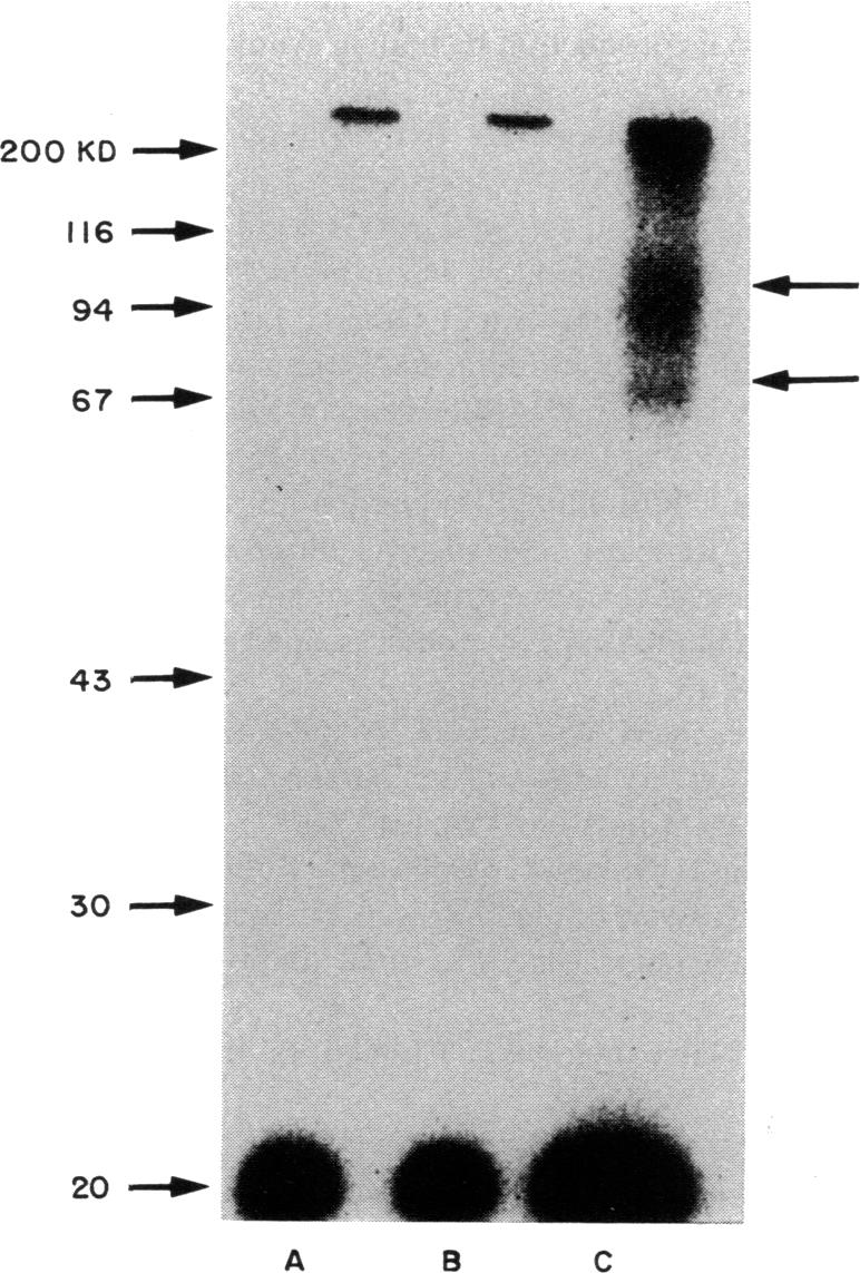
IL-1 possesses pleiotropic properties on various cells and its activity may be stringently regulated in several ways. We have previously reported that both IL-1 and its inhibitory factor are concomitantly released from alveolar macrophages in both healthy subjects and patients with chronic inflammatory lung diseases. An increase in IL-1 activities and a decrease in inhibitory activities are characteristics found in both healthy smokers and patients with interstitial lung diseases. In this study, we further examined the biological properties of IL-1 inhibitory factor. The inhibitor exhibited a dose-dependent specific inhibition of an augmentation by IL-1 of PHA-induced murine thymocyte proliferation, while no inhibition of the augmentation by IL-2, IL-4, IL-6, or tumour necrosis factor (TNF) was found. 125I-labelled IL-1 alpha binding on PHA-stimulated murine thymocytes revealed two types of IL-1 binding sites, 44 sites/cell with a Kd of 2.7 x 10(-10) M and 230 sites/cell with a Kd of 2.5 x 10(-9) M. Alveolar macrophage culture supernatants blocked the binding of labelled IL-1 to the IL-1 receptor in a dose-dependent fashion. Scatchard plot analysis revealed that the inhibitory factor in the supernatants blocked the binding competitively. These results indicate that alveolar macrophages produce a specific IL-1 inhibitory factor, functioning as an IL-1 receptor antagonist.
白细胞介素-1(IL-1)对多种细胞具有多效性,其活性可能通过多种方式受到严格调控。我们之前报道过,在健康受试者和慢性炎症性肺病患者中,IL-1及其抑制因子均从肺泡巨噬细胞中同时释放。IL-1活性增加和抑制活性降低是健康吸烟者和间质性肺病患者共有的特征。在本研究中,我们进一步检测了IL-1抑制因子的生物学特性。该抑制剂对IL-1增强PHA诱导的小鼠胸腺细胞增殖表现出剂量依赖性的特异性抑制,而未发现对IL-2、IL-4、IL-6或肿瘤坏死因子(TNF)增强作用的抑制。用125I标记的IL-1α与PHA刺激的小鼠胸腺细胞结合显示出两种IL-1结合位点,一种是每个细胞44个位点,解离常数(Kd)为2.7×10⁻¹⁰M,另一种是每个细胞230个位点,Kd为2.5×10⁻⁹M。肺泡巨噬细胞培养上清液以剂量依赖性方式阻断标记的IL-1与IL-1受体的结合。Scatchard图分析表明,上清液中的抑制因子竞争性地阻断了这种结合。这些结果表明,肺泡巨噬细胞产生一种特异性IL-1抑制因子,其作用为IL-1受体拮抗剂。