Churchill James D, Tharp Jason A, Wellman Cara L, Sengelaub Dale R, Garraghty Preston E
Department of Psychology, Saint Louis University, St. Louis, Missouri 63103, USA.
BMC Neurosci. 2004 Nov 8;5:43. doi: 10.1186/1471-2202-5-43.
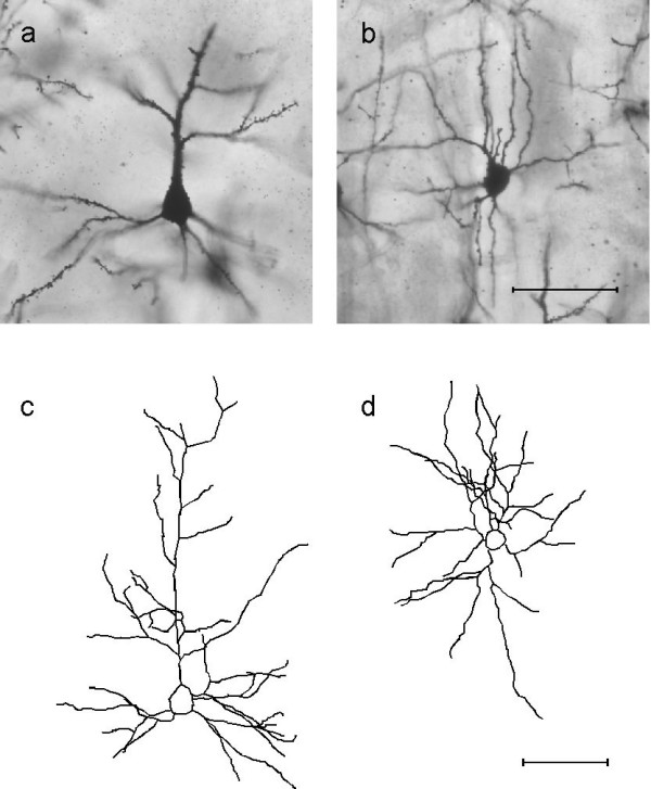
Topographic reorganization of central maps following peripheral nerve injury has been well characterized. Despite extensive documentation of these physiological changes, the underlying anatomical correlates have yet to be fully explored. In this study, we used Golgi impregnation and light microscopy to assess dendritic morphology following denervation of the glabrous hand surface in adult primates.
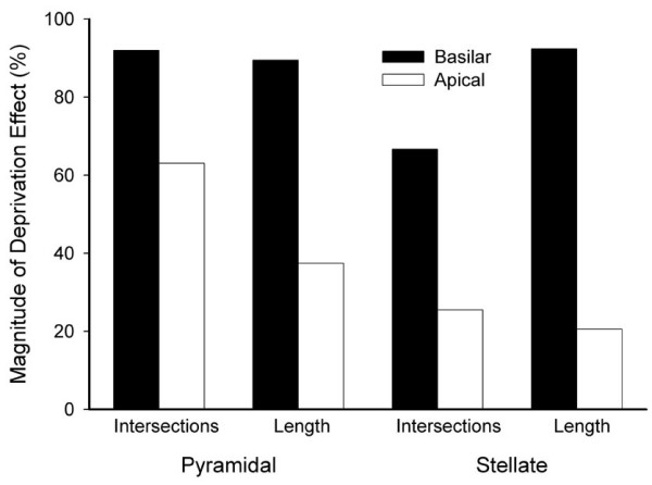
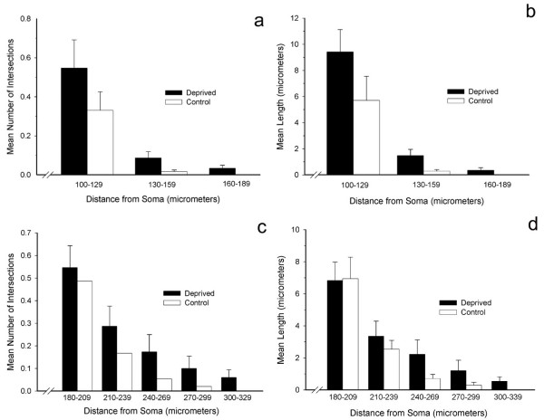
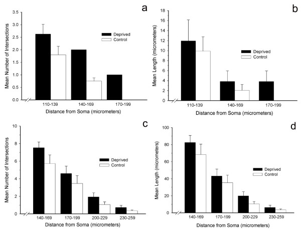
After survival durations that permit complete physiologically-defined reorganization, we find a systematic change in the dendritic arborization pattern of both layer II/III pyramidal and layer IV spiny stellate cells in the contralateral hand region of area 3b, compared to unaffected cortical areas. In general, our analyses indicate a progressive expansion of distal regions of the dendritic arbor with no appreciable changes proximally. This pattern of distal dendritic elaboration occurs for both basilar and apical dendrites.
These observations are consistent with the notion that latent inputs gain expression in reorganized cortex after nerve injury via their influence through contacts with more distally located termination sites.
周围神经损伤后中枢图谱的拓扑重组已得到充分表征。尽管对这些生理变化有大量记录,但潜在的解剖学关联尚未得到充分探索。在本研究中,我们使用高尔基染色法和光学显微镜来评估成年灵长类动物无毛手部表面去神经支配后树突形态。
在允许进行完全生理定义的重组的存活期后,我们发现与未受影响的皮质区域相比,3b区对侧手部区域的II/III层锥体细胞和IV层棘状星状细胞的树突分支模式发生了系统性变化。总体而言,我们的分析表明树突远端区域逐渐扩展,近端没有明显变化。这种远端树突细化模式在基底树突和顶树突中均有出现。
这些观察结果与以下观点一致,即潜在输入在神经损伤后通过与更位于远端的终末位点接触产生影响,从而在重组皮质中获得表达。