• 文献检索
  • 文档翻译
  • 深度研究
  • 学术资讯
  • Suppr Zotero 插件Zotero 插件
  • 邀请有礼
  • 套餐&价格
  • 历史记录
应用&插件
Suppr Zotero 插件Zotero 插件浏览器插件Mac 客户端Windows 客户端微信小程序
定价
高级版会员购买积分包购买API积分包
服务
文献检索文档翻译深度研究API 文档MCP 服务
关于我们
关于 Suppr公司介绍联系我们用户协议隐私条款
关注我们

Suppr 超能文献

核心技术专利:CN118964589B侵权必究
粤ICP备2023148730 号-1Suppr @ 2026

文献检索

告别复杂PubMed语法,用中文像聊天一样搜索,搜遍4000万医学文献。AI智能推荐,让科研检索更轻松。

立即免费搜索

文件翻译

保留排版,准确专业,支持PDF/Word/PPT等文件格式,支持 12+语言互译。

免费翻译文档

深度研究

AI帮你快速写综述,25分钟生成高质量综述,智能提取关键信息,辅助科研写作。

立即免费体验

小角X射线散射(SAXS)在正常乳腺组织与癌性乳腺组织鉴别中的应用。

Application of small angle X-ray scattering (SAXS) for differentiation between normal and cancerous breast tissue.

作者信息

Changizi Vahid, Oghabian Mohammad A, Speller Robert, Sarkar Saeed, Kheradmand Ali Arab

机构信息

Department of Medical Physics, Faculty of Medicine, Tehran University of Medical Sciences, & Research Center for Science & Technology in Medicine, Imam Khomaini Hospital, Bolvare Keshavarz, Tehran, Iran.

出版信息

Int J Med Sci. 2005;2(3):118-21. doi: 10.7150/ijms.2.118. Epub 2005 Jul 5.

DOI:10.7150/ijms.2.118
PMID:16007264
原文链接:https://pmc.ncbi.nlm.nih.gov/articles/PMC1168877/
Abstract

INTRODUCTION

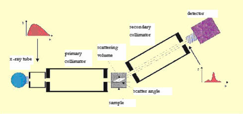
Small angle, between 3 degrees and 10 degrees, X ray scattering is predominantly coherent giving rise to diffraction effects that can be observed as constructive and destructive interferences. These interferences carry information about the molecular structure of the tissue and hence can be used to identify changes that occur due to cancer.

METHOD

In this study an energy dispersive X-ray diffraction method was used. The optimum scattering angle, determined from a series of measurements on adipose breast tissue at several angles from 4 to 7.3 degrees, was found to be 6.5 degrees. Once optimized the system was used to measure the diffraction profiles (corrected scattered intensity versus momentum transfer) of a total of 99 breast tissue samples. The samples were both normal and tumour samples.

RESULTS

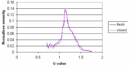
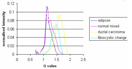
Adipose tissue showed a sharp, high intensity peak at low momentum transfer values of approximately 1.1nm-1. Adipose tissue, mixed tissue (adipose & fibroglandular) and tumor have peaks at different values of momentum transfer that can be used to identify the tissue. Benign and malignant breast tissues can also be differentiated by both peak positions and peak heights. It was also observed that the results were reproducible even after the tissue had been preserved at liquid nitrogen temperatures.

CONCLUSION

We were able to differentiate between normal, benign and malignant breast tissues by using energy dispersive small angle x-ray scattering.

摘要

引言

小角度(3度至10度)X射线散射主要是相干散射,会产生衍射效应,可观察到相长干涉和相消干涉。这些干涉携带了有关组织分子结构的信息,因此可用于识别因癌症而发生的变化。

方法

在本研究中使用了能量色散X射线衍射方法。通过对脂肪乳腺组织在4度至7.3度的几个角度进行一系列测量,确定最佳散射角为6.5度。一旦优化后,该系统用于测量总共99个乳腺组织样本的衍射图谱(校正后的散射强度与动量转移关系)。样本包括正常样本和肿瘤样本。

结果

脂肪组织在约1.1nm-1的低动量转移值处显示出一个尖锐的高强度峰。脂肪组织、混合组织(脂肪与纤维腺组织)和肿瘤在不同的动量转移值处有峰,可用于识别组织。良性和恶性乳腺组织也可通过峰位置和峰高进行区分。还观察到即使在液氮温度下保存组织后,结果仍具有可重复性。

结论

我们能够通过使用能量色散小角度X射线散射来区分正常、良性和恶性乳腺组织。

https://cdn.ncbi.nlm.nih.gov/pmc/blobs/72dc/1168877/2552277324d8/ijmsv02p0118g03.jpg
https://cdn.ncbi.nlm.nih.gov/pmc/blobs/72dc/1168877/c68651731373/ijmsv02p0118g01.jpg
https://cdn.ncbi.nlm.nih.gov/pmc/blobs/72dc/1168877/e9807bb99755/ijmsv02p0118g02.jpg
https://cdn.ncbi.nlm.nih.gov/pmc/blobs/72dc/1168877/2552277324d8/ijmsv02p0118g03.jpg
https://cdn.ncbi.nlm.nih.gov/pmc/blobs/72dc/1168877/c68651731373/ijmsv02p0118g01.jpg
https://cdn.ncbi.nlm.nih.gov/pmc/blobs/72dc/1168877/e9807bb99755/ijmsv02p0118g02.jpg
https://cdn.ncbi.nlm.nih.gov/pmc/blobs/72dc/1168877/2552277324d8/ijmsv02p0118g03.jpg

相似文献

1
Application of small angle X-ray scattering (SAXS) for differentiation between normal and cancerous breast tissue.小角X射线散射(SAXS)在正常乳腺组织与癌性乳腺组织鉴别中的应用。
Int J Med Sci. 2005;2(3):118-21. doi: 10.7150/ijms.2.118. Epub 2005 Jul 5.
2
Application of small-angle X-ray scattering for differentiation among breast tumors.小角X射线散射在乳腺肿瘤鉴别中的应用。
J Med Phys. 2008 Jan;33(1):19-23. doi: 10.4103/0971-6203.39420.
3
Preliminary study of human breast tissue using synchrotron radiation combining WAXS and SAXS techniques.使用同步辐射结合广角X射线散射(WAXS)和小角X射线散射(SAXS)技术对人体乳腺组织进行的初步研究。
Appl Radiat Isot. 2010 Apr-May;68(4-5):799-803. doi: 10.1016/j.apradiso.2009.09.064. Epub 2009 Oct 7.
4
Structural Studies of Epithelial Mesenchymal Transition Breast Tissues.上皮间质转化乳腺组织的结构研究。
Sci Rep. 2020 Feb 6;10(1):1997. doi: 10.1038/s41598-020-58932-5.
5
Analysis of breast cancer by small angle X-ray scattering (SAXS).小角 X 射线散射(SAXS)分析乳腺癌。
Analyst. 2009 Jun;134(6):1077-82. doi: 10.1039/b821434d. Epub 2009 Apr 6.
6
A semianalytic model to extract differential linear scattering coefficients of breast tissue from energy dispersive x-ray diffraction measurements.
Med Phys. 2006 Apr;33(4):959-67. doi: 10.1118/1.2170616.
7
The use of Compton scattering to differentiate between classifications of normal and diseased breast tissue.
Phys Med Biol. 2005 Jul 21;50(14):3337-48. doi: 10.1088/0031-9155/50/14/010. Epub 2005 Jul 6.
8
Human breast cancer in vitro: matching histo-pathology with small-angle x-ray scattering and diffraction enhanced x-ray imaging.人乳腺癌体外研究:组织病理学与小角X射线散射及衍射增强X射线成像的匹配
Phys Med Biol. 2005 Jul 7;50(13):2991-3006. doi: 10.1088/0031-9155/50/13/002. Epub 2005 Jun 8.
9
WAXS fat subtraction model to estimate differential linear scattering coefficients of fatless breast tissue: phantom materials evaluation.用于估计无脂肪乳腺组织微分线性散射系数的小角X射线散射脂肪减法模型:体模材料评估
Med Phys. 2014 May;41(5):053501. doi: 10.1118/1.4870982.
10
Monte Carlo simulation of x-ray scattering for quantitative characterization of breast cancer.用于乳腺癌定量表征的X射线散射蒙特卡罗模拟
Phys Med Biol. 2009 Jun 21;54(12):3773-84. doi: 10.1088/0031-9155/54/12/011. Epub 2009 May 28.

引用本文的文献

1
Structural Studies of Epithelial Mesenchymal Transition Breast Tissues.上皮间质转化乳腺组织的结构研究。
Sci Rep. 2020 Feb 6;10(1):1997. doi: 10.1038/s41598-020-58932-5.
2
Model predictions for the wide-angle x-ray scatter signals of healthy and malignant breast duct biopsies.健康和恶性乳腺导管活检的广角X射线散射信号的模型预测。
J Med Imaging (Bellingham). 2015 Oct;2(4):043502. doi: 10.1117/1.JMI.2.4.043502. Epub 2015 Oct 23.
3
Small-angle X-ray scattering method to characterize molecular interactions: Proof of concept.

本文引用的文献

1
Breast cancer diagnosis using scattered X-rays.利用散射X射线进行乳腺癌诊断。
J Synchrotron Radiat. 2000 Sep 1;7(Pt 5):348-52. doi: 10.1107/S0909049500009973.
2
Small-angle x-ray scattering studies of human breast tissue samples.人类乳腺组织样本的小角X射线散射研究。
Phys Med Biol. 2002 Feb 21;47(4):577-92. doi: 10.1088/0031-9155/47/4/303.
3
X-ray scatter signatures for normal and neoplastic breast tissues.正常和肿瘤性乳腺组织的X射线散射特征。
用于表征分子相互作用的小角X射线散射方法:概念验证。
Sci Rep. 2015 Jul 10;5:12085. doi: 10.1038/srep12085.
4
Modern breast cancer detection: a technological review.现代乳腺癌检测:技术综述
Int J Biomed Imaging. 2009;2009:902326. doi: 10.1155/2009/902326. Epub 2009 Dec 28.
5
Application of small-angle X-ray scattering for differentiation among breast tumors.小角X射线散射在乳腺肿瘤鉴别中的应用。
J Med Phys. 2008 Jan;33(1):19-23. doi: 10.4103/0971-6203.39420.
Phys Med Biol. 1999 Jul;44(7):1791-802. doi: 10.1088/0031-9155/44/7/316.
4
X-ray diffraction measurements of some plastic materials and body tissues.
Med Phys. 1987 Jul-Aug;14(4):526-32. doi: 10.1118/1.596143.
5
Estimates of the worldwide frequency of sixteen major cancers in 1980.1980年全球16种主要癌症发病率的估计数据。
Int J Cancer. 1988 Feb 15;41(2):184-97. doi: 10.1002/ijc.2910410205.
6
Measurement of small-angle photon scattering for some breast tissues and tissue substitute materials.
Phys Med Biol. 1991 Jan;36(1):7-18. doi: 10.1088/0031-9155/36/1/002.