Sharma Rakesh, Kline Richard P, Wu Ed X, Katz Jose K
Department of Medicine, Columbia University, New York, NY 10032, USA.
Cancer Cell Int. 2005 Aug 3;5:26. doi: 10.1186/1475-2867-5-26.
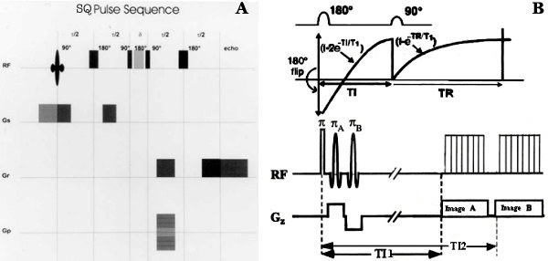
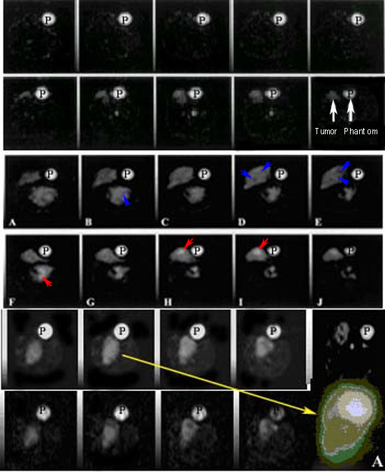
Sodium weighted images can indicate sodium signal intensities from different features in the tumor before and 24 hours following administration of Taxotere.
To evaluate the association of in vivo intracellular sodium magnetic resonance image intensities with immuno-biomarkers and histopathological features to monitor the early tumor response to Taxotere chemotherapy in Methyl-Nitroso-Urea induced rat xenograft breast tumors.
Methyl-Nitroso-Urea (MNU) induced rat xenograft breast tumors were imaged for sodium MRI and compared with tumor histology, immunostaining after 24 hours chemotherapy.
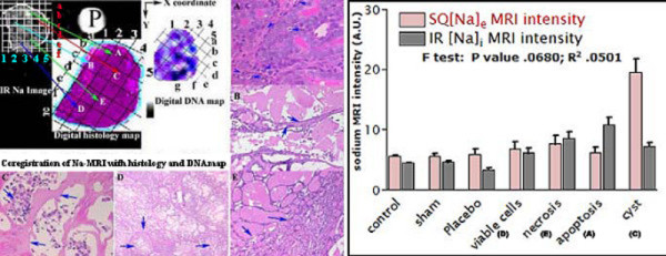
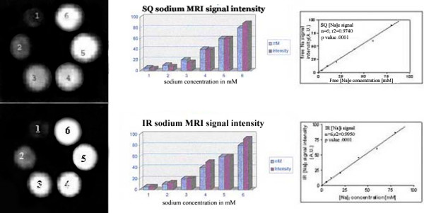
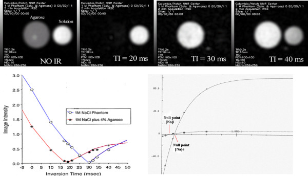
Sodium MRI signal intensities represented sodium concentrations. Excised tumor histological sections showed different in vitro histological end points i.e. single strand DNA content of cell nuclei during cell cycle (G1/S-G2/M), distinct S or M histograms (Feulgen labeling to nuclear DNA content by CAS 200), mitotic figures and apoptosis at different locations of breast tumors. Necrosis and cystic fluid appeared gray on intracellular (IC) sodium images while apoptosis rich regions appeared brighter on IC sodium images. After 24 hours Taxotere-treated tumors showed lower 'IC/EC ratio' of viable cells (65-76%) with higher mitotic index; apoptotic tumor cells at high risk due to cytotoxicity (>70% with high apoptotic index); reduced proliferation index (270 vs 120 per high power field) associated with enhanced IC sodium in vivo MR image intensities and decreased tumor size (3%; p < 0.001; n = 16) than that of pre-treated tumors. IC-Na MR signal intensities possibly indicated Taxotere chemosensitivity response in vivo associated with apoptosis and different pre-malignant features within 24 hours of exposure of cancer cells to anti-neoplastic Taxotere drug.
Sodium MRI imaging may be used as in vivo rapid drug monitoring method to evaluate Taxotere chemosensitivity response associated with neoplasia, apoptosis and tumor histology features.
钠加权图像可显示在给予多西他赛后24小时内及之前肿瘤不同特征的钠信号强度。
评估体内细胞内钠磁共振图像强度与免疫生物标志物及组织病理学特征之间的关联,以监测甲基亚硝基脲诱导的大鼠异种移植乳腺肿瘤对多西他赛化疗的早期肿瘤反应。
对甲基亚硝基脲(MNU)诱导的大鼠异种移植乳腺肿瘤进行钠MRI成像,并与肿瘤组织学、化疗24小时后的免疫染色进行比较。
钠MRI信号强度代表钠浓度。切除的肿瘤组织学切片显示出不同的体外组织学终点,即细胞周期(G1/S-G2/M)期间细胞核的单链DNA含量、不同的S或M直方图(通过CAS 200对核DNA含量进行福尔根标记)、乳腺肿瘤不同位置的有丝分裂图像和凋亡。坏死和囊液在细胞内(IC)钠图像上呈灰色,而富含凋亡区域在IC钠图像上显得更亮。多西他赛治疗24小时后,存活细胞的“IC/EC比率”较低(65-76%),有丝分裂指数较高;由于细胞毒性,凋亡肿瘤细胞处于高风险(凋亡指数高时>70%);增殖指数降低(每高倍视野270对120),与体内IC钠MR图像强度增强和肿瘤大小减小(3%;p<0.001;n=16)相关,与治疗前肿瘤相比。IC-Na MR信号强度可能表明在癌细胞暴露于抗肿瘤多西他赛药物24小时内,体内多西他赛的化学敏感性反应与凋亡及不同的癌前特征相关。
钠MRI成像可作为一种体内快速药物监测方法,以评估与肿瘤形成、凋亡和肿瘤组织学特征相关的多西他赛化学敏感性反应。