Kwong K K, Belliveau J W, Chesler D A, Goldberg I E, Weisskoff R M, Poncelet B P, Kennedy D N, Hoppel B E, Cohen M S, Turner R
Department of Radiology, Massachusetts General Hospital, Charlestown.
Proc Natl Acad Sci U S A. 1992 Jun 15;89(12):5675-9. doi: 10.1073/pnas.89.12.5675.
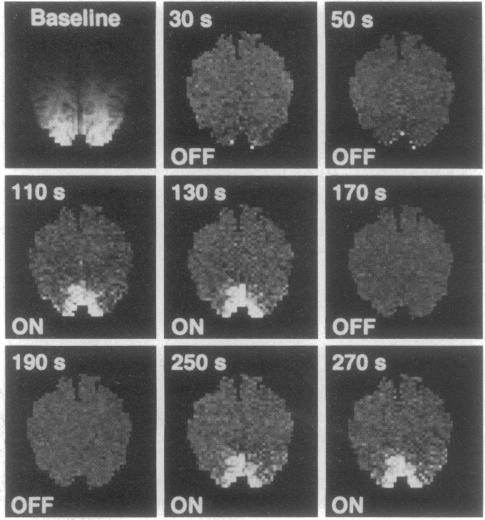
Neuronal activity causes local changes in cerebral blood flow, blood volume, and blood oxygenation. Magnetic resonance imaging (MRI) techniques sensitive to changes in cerebral blood flow and blood oxygenation were developed by high-speed echo planar imaging. These techniques were used to obtain completely noninvasive tomographic maps of human brain activity, by using visual and motor stimulus paradigms. Changes in blood oxygenation were detected by using a gradient echo (GE) imaging sequence sensitive to the paramagnetic state of deoxygenated hemoglobin. Blood flow changes were evaluated by a spin-echo inversion recovery (IR), tissue relaxation parameter T1-sensitive pulse sequence. A series of images were acquired continuously with the same imaging pulse sequence (either GE or IR) during task activation. Cine display of subtraction images (activated minus baseline) directly demonstrates activity-induced changes in brain MR signal observed at a temporal resolution of seconds. During 8-Hz patterned-flash photic stimulation, a significant increase in signal intensity (paired t test; P less than 0.001) of 1.8% +/- 0.8% (GE) and 1.8% +/- 0.9% (IR) was observed in the primary visual cortex (V1) of seven normal volunteers. The mean rise-time constant of the signal change was 4.4 +/- 2.2 s for the GE images and 8.9 +/- 2.8 s for the IR images. The stimulation frequency dependence of visual activation agrees with previous positron emission tomography observations, with the largest MR signal response occurring at 8 Hz. Similar signal changes were observed within the human primary motor cortex (M1) during a hand squeezing task and in animal models of increased blood flow by hypercapnia. By using intrinsic blood-tissue contrast, functional MRI opens a spatial-temporal window onto individual brain physiology.
神经元活动会引起脑血流量、血容量和血液氧合的局部变化。对脑血流量和血液氧合变化敏感的磁共振成像(MRI)技术是通过高速回波平面成像技术开发出来的。这些技术被用于通过视觉和运动刺激范式来获取人类大脑活动的完全无创断层图像。通过使用对脱氧血红蛋白的顺磁状态敏感的梯度回波(GE)成像序列来检测血液氧合的变化。通过自旋回波反转恢复(IR)、对组织弛豫参数T1敏感的脉冲序列来评估血流量变化。在任务激活期间,用相同的成像脉冲序列(GE或IR)连续采集一系列图像。减法图像(激活图像减去基线图像)的电影显示直接展示了在秒级时间分辨率下观察到的活动诱导的脑磁共振信号变化。在8赫兹模式闪光光刺激期间,在7名正常志愿者的初级视觉皮层(V1)中观察到信号强度显著增加(配对t检验;P小于0.001),GE序列为1.8%±0.8%,IR序列为1.8%±0.9%。GE图像信号变化的平均上升时间常数为4.4±2.2秒,IR图像为8.9±2.8秒。视觉激活的刺激频率依赖性与先前的正电子发射断层扫描观察结果一致,最大的磁共振信号反应出现在8赫兹。在手部挤压任务期间,在人类初级运动皮层(M1)内以及在高碳酸血症导致血流量增加的动物模型中也观察到了类似的信号变化。通过利用内在的血液-组织对比,功能磁共振成像为个体脑生理学打开了一个时空窗口。