Chung Myung Jin, Lee Kyung Soo, Koh Won-Jung, Lee Ju Hyun, Kim Tae Sung, Kwon O Jung, Kim Seonwoo
Department of Radiology and Center for Imaging Science, Samsung Medical Center, Sungkyunkwan University School of Medicine, Seoul, Korea.
J Korean Med Sci. 2005 Oct;20(5):777-83. doi: 10.3346/jkms.2005.20.5.777.
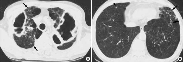
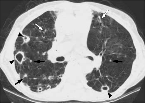
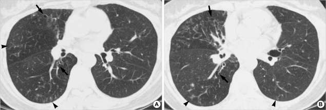
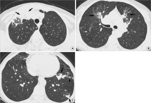
We aimed to compare the CT findings of nontuberculous mycobacterial pulmonary diseases caused by Mycobacterium avium-intracellulare complex (MAC) and Mycobacterium abscessus. Two chest radiologists analyzed retrospectively the thin-section CT findings of 51 patients with MAC and 36 with M. abscessus infection in terms of patterns and forms of lung lesions. No significant difference was found between MAC and M. abscessus infection in the presence of small nodules, tree-in-bud pattern, and bronchiectasis. However, lobar volume decrease (p=0.001), nodule (p=0.018), airspace consolidation (p=0.047) and thin-walled cavity (p=0.009) were more frequently observed in MAC infection. The upper lobe cavitary form was more frequent in the MAC (19 of 51 patients, 37%) group than M. abscessus (5 of 36, 14%) (p=0.029), whereas the nodular bronchiectatic form was more frequent in the M. abscessus group ([29 of 36, 81%] vs. [27 of 51, 53%] in MAC) (p=0.012). In conclusion, there is considerable overlap in common CT findings of MAC and M. abscessus pulmonary infection; however, lobar volume loss, nodule, airspace consolidation, and thin-walled cavity are more frequently seen in MAC than M. abscessus infection.
我们旨在比较由鸟分枝杆菌复合群(MAC)和脓肿分枝杆菌引起的非结核分枝杆菌肺病的CT表现。两位胸部放射科医生回顾性分析了51例MAC感染患者和36例脓肿分枝杆菌感染患者的薄层CT表现,包括肺部病变的模式和形态。MAC感染和脓肿分枝杆菌感染在小结节、树芽征和支气管扩张的出现方面未发现显著差异。然而,MAC感染中更常观察到肺叶体积缩小(p = 0.001)、结节(p = 0.018)、气腔实变(p = 0.047)和薄壁空洞(p = 0.009)。MAC组(51例患者中的19例,37%)上叶空洞型比脓肿分枝杆菌组(36例中的5例,14%)更常见(p = 0.029),而结节性支气管扩张型在脓肿分枝杆菌组中更常见([36例中的29例,81%] vs. MAC组中的[51例中的27例,53%])(p = 0.012)。总之,MAC和脓肿分枝杆菌肺部感染的常见CT表现有相当大的重叠;然而,MAC感染比脓肿分枝杆菌感染更常出现肺叶体积缩小、结节、气腔实变和薄壁空洞。