Summers Ronald M, Yao Jianhua, Pickhardt Perry J, Franaszek Marek, Bitter Ingmar, Brickman Daniel, Krishna Vamsi, Choi J Richard
Diagnostic Radiology Department, Warren Grant Magnuson Clinical Center, National Institutes of Health, Bethesda, Maryland 20892-1182, USA.
Gastroenterology. 2005 Dec;129(6):1832-44. doi: 10.1053/j.gastro.2005.08.054.
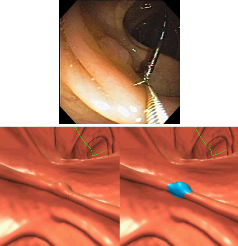
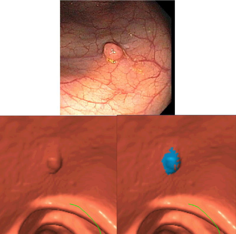
BACKGROUND & AIMS: The sensitivity of computed tomographic (CT) virtual colonoscopy (CT colonography) for detecting polyps varies widely in recently reported large clinical trials. Our objective was to determine whether a computer program is as sensitive as optical colonoscopy for the detection of adenomatous colonic polyps on CT virtual colonoscopy.
The data set was a cohort of 1186 screening patients at 3 medical centers. All patients underwent same-day virtual and optical colonoscopy. Our enhanced gold standard combined segmental unblinded optical colonoscopy and retrospective identification of precise polyp locations. The data were randomized into separate training (n = 394) and test (n = 792) sets for analysis by a computer-aided polyp detection (CAD) program.
For the test set, per-polyp and per-patient sensitivities for CAD were both 89.3% (25/28; 95% confidence interval, 71.8%-97.7%) for detecting retrospectively identifiable adenomatous polyps at least 1 cm in size. The false-positive rate was 2.1 (95% confidence interval, 2.0-2.2) false polyps per patient. Both carcinomas were detected by CAD at a false-positive rate of 0.7 per patient; only 1 of 2 was detected by optical colonoscopy before segmental unblinding. At both 8-mm and 10-mm adenoma size thresholds, the per-patient sensitivities of CAD were not significantly different from those of optical colonoscopy before segmental unblinding.
The per-patient sensitivity of CT virtual colonoscopy CAD in an asymptomatic screening population is comparable to that of optical colonoscopy for adenomas > or = 8 mm and is generalizable to new CT virtual colonoscopy data.
在最近报道的大型临床试验中,计算机断层扫描(CT)虚拟结肠镜检查(CT结肠成像)检测息肉的敏感性差异很大。我们的目的是确定计算机程序在CT虚拟结肠镜检查中检测结肠腺瘤性息肉时是否与光学结肠镜检查一样敏感。
数据集来自3个医疗中心的1186名筛查患者队列。所有患者均在同一天接受了虚拟结肠镜检查和光学结肠镜检查。我们改进的金标准结合了分段非盲法光学结肠镜检查和息肉精确位置的回顾性识别。数据被随机分为单独的训练集(n = 394)和测试集(n = 792),由计算机辅助息肉检测(CAD)程序进行分析。
对于测试集,CAD检测至少1 cm大小的回顾性可识别腺瘤性息肉的息肉敏感性和患者敏感性均为89.3%(25/28;95%置信区间,71.8%-97.7%)。假阳性率为每位患者2.1个(95%置信区间,2.0-2.2)假息肉。CAD检测到了两种癌,假阳性率为每位患者0.7;在分段非盲法之前,光学结肠镜检查仅检测到了2例中的1例。在8 mm和10 mm腺瘤大小阈值时,CAD的患者敏感性与分段非盲法之前的光学结肠镜检查相比无显著差异。
在无症状筛查人群中,CT虚拟结肠镜检查CAD的患者敏感性与光学结肠镜检查对≥8 mm腺瘤的敏感性相当,并且可推广到新的CT虚拟结肠镜检查数据。