Department of Plant Pathology, University of Minnesota, St. Paul, Minnesota 55108; Repligen-Sandoz Research Corp., Lexington, Massachusetts 02173 ; and Northern Regional Research Center, Agricultural Research Service, U.S. Department of Agriculture, Peoria, Illinois 61604.
Appl Environ Microbiol. 1989 Jun;55(6):1457-65. doi: 10.1128/aem.55.6.1457-1465.1989.
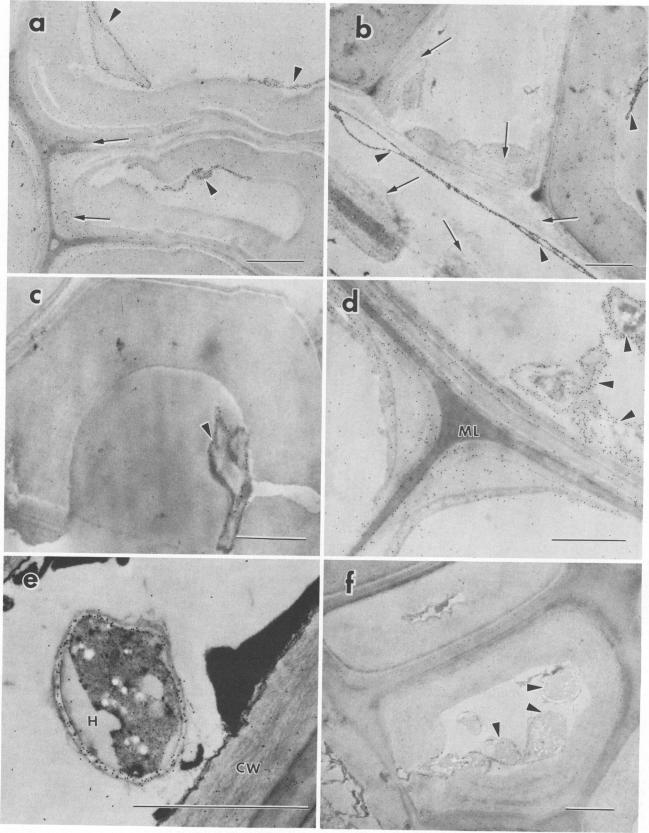
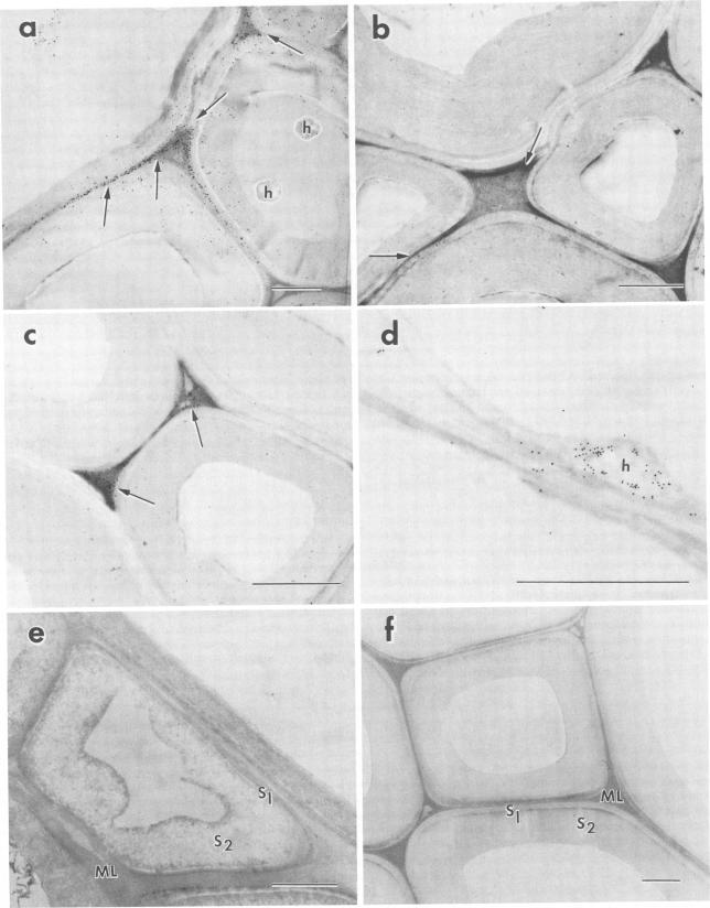
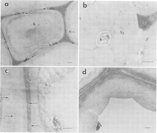
The white rot fungi used in this study caused two different forms of degradation. Phanerochaete chrysosporium, strain BKM-F-1767, and Phellinus pini caused a preferential removal of lignin from birch wood, whereas Trametes (Coriolus) versicolor caused a nonselective attack of all cell wall components. Use of polyclonal antisera to H8 lignin peroxidase and monoclonal antisera to H2 lignin peroxidase followed by immunogold labeling with protein A-gold or protein G-gold, respectively, showed lignin peroxidase extra-and intracellularly to fungal hyphae and within the delignified cell walls after 12 weeks of laboratory decay. Lignin peroxidase was localized at sites within the cell wall where electron-dense areas of the lignified cell wall layers remained. In wood decayed by Trametes versicolor, lignin peroxidase was located primarily along the surface of eroded cell walls. No lignin peroxidase was evident in brown-rotted wood, but slight labeling occurred within hyphal cells. Use of polyclonal antisera to xylanase followed by immunogold labeling showed intense labeling on fungal hyphae and surrounding slime layers and within the woody cell wall, where evidence of degradation was apparent. Colloidal-gold-labeled xylanase was prevalent in wood decayed by all fungi used in this study. Areas of the wood with early stages of cell wall decay had the greatest concentration of gold particles, while little labeling occurred in cells in advanced stages of decay by brown or white rot fungi.
本研究中使用的白腐真菌导致了两种不同形式的降解。栓菌(Phanerochaete chrysosporium)菌株 BKM-F-1767 和松栓菌(Phellinus pini)优先去除桦木中的木质素,而彩绒革盖菌(Trametes (Coriolus) versicolor)则对所有细胞壁成分进行非选择性攻击。使用多克隆抗血清针对 H8 木质素过氧化物酶和单克隆抗血清针对 H2 木质素过氧化物酶,然后分别用蛋白 A-金或蛋白 G-金进行免疫金标记,显示木质素过氧化物酶在真菌菌丝的细胞外和细胞内以及木质素脱除细胞壁内。木质素过氧化物酶定位于细胞壁内的木质素层电子致密区仍然存在的部位。在彩绒革盖菌腐朽的木材中,木质素过氧化物酶主要位于侵蚀细胞壁的表面。在褐腐木材中没有明显的木质素过氧化物酶,但在菌丝细胞内有轻微的标记。使用针对木聚糖酶的多克隆抗血清,然后进行免疫金标记,显示在真菌菌丝和周围的黏液层以及木质细胞壁内有强烈的标记,明显有降解的迹象。胶体金标记的木聚糖酶在本研究中使用的所有真菌腐朽的木材中都很普遍。细胞壁早期降解的区域金颗粒浓度最高,而在褐腐或白腐真菌晚期降解的细胞中几乎没有标记。