Baehni P C, Song M, McCulloch C A, Ellen R P
School of Dental Medicine, University of Geneva, Switzerland.
Infect Immun. 1992 Aug;60(8):3360-8. doi: 10.1128/iai.60.8.3360-3368.1992.
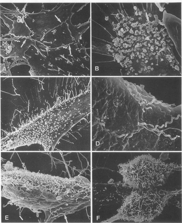
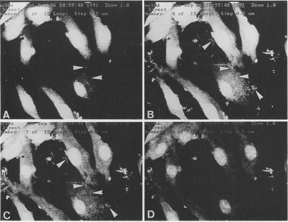
Spirochetes are associated with destructive periodontal diseases, and one cultivatable oral species, Treponema denticola, binds to mammalian cells and perturbs metabolism. To evaluate the cytoskeletal responses and attachment functions of human gingival fibroblasts (HGF) exposed to T. denticola, monolayers of HGF were incubated with T. denticola strains ATCC 35405, e, and e' in serum-free medium. HGF retracted pseudopods, rounded up, and ultimately detached from the substratum. Scanning electron microscopy showed spirochetes in close contact with HGF surfaces; occasionally, bacteria were partially submerged between folds in the HGF membrane. Blebbing and numerous microvilli formed on the cell surface as the HGF retracted. By confocal microscopy, spirochetes were detected in contact with the HGF surface but were never found on the ventral surface of fibroblasts between the substratum and cell. Morphological alterations were associated with and preceded by actin assembly, as measured by microscopic fluorimetry: there was a 263% increase in actin fluorescence over controls within 30 min. Detachment of fibroblasts from the substratum was related to incubation time and was dependent on the concentration of T. denticola. Detachment was observed for all strains tested and was also dependent on the viability of T. denticola: UV light, heat, and metronidazole treatment markedly reduced the HGF detachment response. Detachment was also significantly reduced by the protease inhibitor phenylmethylsulfonyl fluoride. HGF viability was not significantly affected by coincubation with spirochetes, as measured by lactate dehydrogenase release. Thus, T. denticola induces rapid cytoskeletal remodelling followed by cell detachment, which might be stimulated by a bacterially associated protease but is not likely directly mediated by proteolytic degradation at the cell-substratum adhesive contact points.
螺旋体与破坏性牙周疾病有关,一种可培养的口腔菌种——齿垢密螺旋体,能与哺乳动物细胞结合并扰乱新陈代谢。为了评估暴露于齿垢密螺旋体的人牙龈成纤维细胞(HGF)的细胞骨架反应和附着功能,将HGF单层细胞在无血清培养基中与齿垢密螺旋体菌株ATCC 35405、e和e'一起孵育。HGF缩回伪足,变圆,最终从基质上脱离。扫描电子显微镜显示螺旋体与HGF表面紧密接触;偶尔,细菌部分淹没在HGF膜的褶皱之间。随着HGF缩回,细胞表面形成了泡状突起和许多微绒毛。通过共聚焦显微镜观察,发现螺旋体与HGF表面接触,但在成纤维细胞位于基质和细胞之间的腹侧表面上从未发现。通过显微荧光测定法测量,形态学改变与肌动蛋白组装相关且先于肌动蛋白组装:30分钟内肌动蛋白荧光比对照增加了263%。成纤维细胞从基质上的脱离与孵育时间有关,并且取决于齿垢密螺旋体的浓度。在所测试的所有菌株中均观察到脱离,并且还取决于齿垢密螺旋体的活力:紫外线、加热和甲硝唑处理显著降低了HGF的脱离反应。蛋白酶抑制剂苯甲基磺酰氟也显著降低了脱离。通过乳酸脱氢酶释放量测定,与螺旋体共孵育对HGF活力没有显著影响。因此,齿垢密螺旋体诱导快速的细胞骨架重塑,随后细胞脱离,这可能由细菌相关的蛋白酶刺激,但不太可能直接由细胞-基质粘附接触点处的蛋白水解降解介导。