Binesh Nader, Huda Amir, Thomas M Albert, Wyckoff Nathaniel, Bugbee Mary, Han Steven, Rasgon Natalie, Davanzo Pablo, Sayre James, Guze Barry, Martin Paul, Fawzy Fawzy
Department of Radiological Sciences, University of California, Los Angeles, California 90095, USA.
J Appl Clin Med Phys. 2006 Winter;7(1):86-96. doi: 10.1120/jacmp.v7i1.2151. Epub 2006 Feb 15.
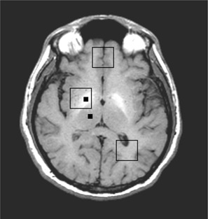
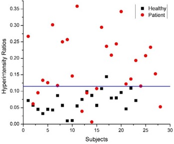
Hepatic encephalopathy (HE) is normally diagnosed by neuropsychological (NP) tests, which are not very specific and do not reveal the underlying pathology. Magnetic resonance imaging (MRI) and spectroscopy (MRS) of the brain offer alternative and possibly more specific markers for HE. These methods were applied in conjunction with NP testing in order to determine their usefulness in the identification of HE and to understand the pathogenesis of HE more clearly. MR imaging and spectroscopy examinations, in addition to a battery of 15 NP tests, were administered to investigate 31 patients awaiting liver transplantation and 23 healthy controls. MR image intensities from the globus pallidus region were calculated and normalized to those of the thalamus. Absolute concentrations and ratios with respect to creatine (Cr) of several metabolites were computed from MR spectra. The MR data were correlated with the results of NP tests. The patients showed impairment in NP tests of attention and visuospatial and verbal fluency. In T1-weighted MRI, the relative intensity of the globus pallidus with respect to that of the thalamus region was significantly elevated in patients and correlated(negatively) with three NP tests (Hooper, FAS, and Trails B). The absolute concentrations of myo-inositol (mI) and choline (Ch) were significantly reduced in three brain regions. In addition, the absolute concentrations of glutamine (Gln) and combined glutamate and glutamine (Glx) were increased in all three locations, with Gln increase being significant in all areas while that of Glx only in the occipital white matter. In summary, this study partially confirms a hypothesized mechanism of HE pathogenesis, an increased synthesis of glutamine by brain glutamate in astrocytes due to excessive blood ammonia, followed by a compensatory loss of myo-inositol to maintain astrocyte volume homeostasis. It also indicates that the hyperintensity observed in globus pallidus could be used as complementary to the NP test scores in evaluating the mental health of HE patients.
肝性脑病(HE)通常通过神经心理学(NP)测试来诊断,这些测试特异性不强,无法揭示潜在的病理情况。脑部磁共振成像(MRI)和波谱分析(MRS)为肝性脑病提供了替代性的、可能更具特异性的标志物。将这些方法与NP测试结合应用,以确定它们在肝性脑病识别中的有用性,并更清楚地了解肝性脑病的发病机制。除了一系列15项NP测试外,还对31名等待肝移植的患者和23名健康对照者进行了MR成像和波谱检查。计算苍白球区域的MR图像强度,并将其与丘脑的强度进行归一化。从MR波谱中计算出几种代谢物相对于肌酸(Cr)的绝对浓度和比率。将MR数据与NP测试结果进行关联。患者在注意力、视觉空间和语言流畅性的NP测试中表现出受损。在T1加权MRI中,患者苍白球相对于丘脑区域的相对强度显著升高,并且与三项NP测试(胡珀测试、FAS测试和连线测验B)(呈负)相关。在三个脑区中,肌醇(mI)和胆碱(Ch)的绝对浓度显著降低。此外,在所有三个位置,谷氨酰胺(Gln)以及谷氨酸和谷氨酰胺总和(Glx)的绝对浓度均升高,其中Gln在所有区域均显著升高,而Glx仅在枕叶白质中显著升高。总之,本研究部分证实了肝性脑病发病机制的一个假设机制,即由于血氨过多,星形胶质细胞中的脑谷氨酸合成谷氨酰胺增加,随后肌醇代偿性丢失以维持星形胶质细胞体积的稳态。它还表明,苍白球中观察到的高信号强度可作为NP测试分数的补充,用于评估肝性脑病患者的心理健康。