Nikonov Sergei S, Kholodenko Roman, Lem Janis, Pugh Edward N
FM Kirby Center for Molecular Ophthalmology, Department of Ophthalmology, School of Medicine, University of Pennsylvania, Philadelphia, 19104, USA.
J Gen Physiol. 2006 Apr;127(4):359-74. doi: 10.1085/jgp.200609490.
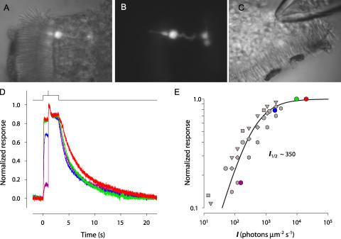
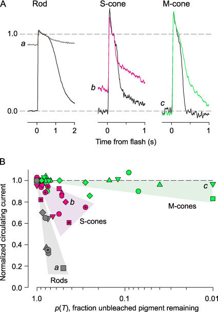
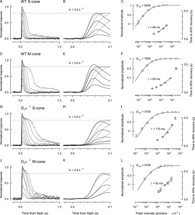
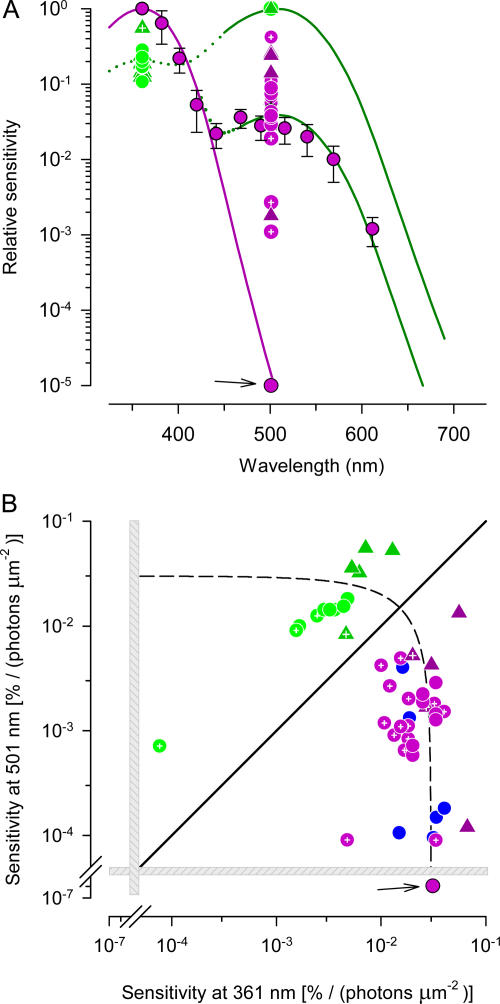
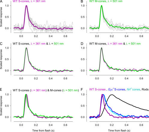
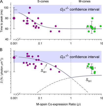
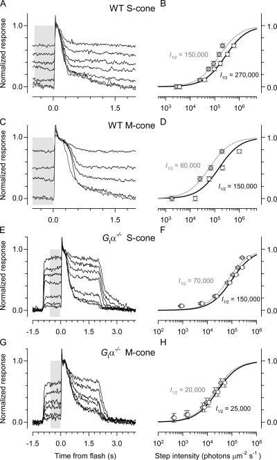
Cone cells constitute only 3% of the photoreceptors of the wild-type (WT) mouse. While mouse rods have been thoroughly investigated with suction pipette recordings of their outer segment membrane currents, to date no recordings from WT cones have been published, likely because of the rarity of cones and the fragility of their outer segments. Recently, we characterized the photoreceptors of Nrl(-/-) mice, using suction pipette recordings from their "inner segments" (perinuclear region), and found them to be cones. Here we report the use of this same method to record for the first time the responses of single cones of WT mice, and of mice lacking the alpha-subunit of the G-protein transducin (G(t)alpha(-/-)), a loss that renders them functionally rodless. Most cones were found to functionally co-express both S- (lambda(max) = 360 nm) and M- (lambda(max) = 508 nm) cone opsins and to be maximally sensitive at 360 nm ("S-cones"); nonetheless, all cones from the dorsal retina were found to be maximally sensitive at 508 nm ("M-cones"). The dim-flash response kinetics and absolute sensitivity of S- and M-cones were very similar and not dependent on which of the coexpressed cone opsins drove transduction; the time to peak of the dim-flash response was approximately 70 ms, and approximately 0.2% of the circulating current was suppressed per photoisomerization. Amplification in WT cones (A approximately 4 s(-2)) was found to be about twofold lower than in rods (A approximately 8 s(-2)). Mouse M-cones maintained their circulating current at very nearly the dark adapted level even when >90% of their M-opsin was bleached. S-cones were less tolerant to bleached S-opsin than M-cones to bleached M-opsin, but still far more tolerant than mouse rods to bleached rhodopsin, which exhibit persistent suppression of nearly 50% of their circulating current following a 20% bleach. Thus, the three types of mouse opsin appear distinctive in the degree to which their bleached, unregenerated opsins generate "dark light."
视锥细胞仅占野生型(WT)小鼠光感受器的3%。虽然已通过对小鼠视杆细胞外段膜电流的吸管记录对其进行了深入研究,但迄今为止,尚未发表过对野生型视锥细胞的记录,这可能是因为视锥细胞稀少且其外段脆弱。最近,我们利用对“内段”(核周区域)的吸管记录对Nrl基因敲除(Nrl(-/-))小鼠的光感受器进行了表征,发现它们是视锥细胞。在此,我们报告首次使用相同方法记录野生型小鼠以及缺乏G蛋白转导素α亚基(G(t)alpha(-/-))的小鼠单个视锥细胞的反应,这种缺失使它们在功能上无视杆细胞。发现大多数视锥细胞在功能上共表达S型(λmax = 360 nm)和M型(λmax = 508 nm)视锥视蛋白,并且在360 nm处具有最大敏感性(“S视锥细胞”);尽管如此,发现来自视网膜背侧的所有视锥细胞在508 nm处具有最大敏感性(“M视锥细胞”)。S视锥细胞和M视锥细胞的暗闪光反应动力学和绝对敏感性非常相似,并且不依赖于共表达的视锥视蛋白中哪一种驱动转导;暗闪光反应的峰值时间约为70毫秒,每次光异构化约0.2%的循环电流被抑制。发现野生型视锥细胞中的放大倍数(A约为4 s(-2))比视杆细胞中的放大倍数(A约为8 s(-2))低约两倍。即使当超过90%的M视蛋白被漂白时,小鼠M视锥细胞仍将其循环电流维持在非常接近暗适应水平。S视锥细胞比M视锥细胞对漂白的S视蛋白的耐受性低,但仍比小鼠视杆细胞对漂白的视紫红质的耐受性高得多,在20%漂白后,视紫红质会持续抑制近50%的循环电流。因此,三种类型的小鼠视蛋白在其漂白的、未再生的视蛋白产生“暗光”的程度上似乎有所不同。