• 文献检索
  • 文档翻译
  • 深度研究
  • 学术资讯
  • Suppr Zotero 插件Zotero 插件
  • 邀请有礼
  • 套餐&价格
  • 历史记录
应用&插件
Suppr Zotero 插件Zotero 插件浏览器插件Mac 客户端Windows 客户端微信小程序
定价
高级版会员购买积分包购买API积分包
服务
文献检索文档翻译深度研究API 文档MCP 服务
关于我们
关于 Suppr公司介绍联系我们用户协议隐私条款
关注我们

Suppr 超能文献

核心技术专利:CN118964589B侵权必究
粤ICP备2023148730 号-1Suppr @ 2026

文献检索

告别复杂PubMed语法,用中文像聊天一样搜索,搜遍4000万医学文献。AI智能推荐,让科研检索更轻松。

立即免费搜索

文件翻译

保留排版,准确专业,支持PDF/Word/PPT等文件格式,支持 12+语言互译。

免费翻译文档

深度研究

AI帮你快速写综述,25分钟生成高质量综述,智能提取关键信息,辅助科研写作。

立即免费体验

不同染色法对眼部微孢子虫病的组织病理学评估。

Histopathological evaluation of ocular microsporidiosis by different stains.

作者信息

Joseph Joveeta, Vemuganti Geeta K, Garg Prashant, Sharma Savitri

机构信息

Jhaveri Microbiology Centre, Hyderabad Eye Research Foundation, L.V.Prasad Eye Institute, L.V.Prasad Marg, Banjara Hills, Hyderabad-500 034, India.

出版信息

BMC Clin Pathol. 2006 Jun 23;6:6. doi: 10.1186/1472-6890-6-6.

DOI:10.1186/1472-6890-6-6
PMID:16792818
原文链接:https://pmc.ncbi.nlm.nih.gov/articles/PMC1513581/
Abstract

BACKGROUND

There is limited data on comparing stains in the detection of microsporidia in corneal biopsies. Hence we wanted to evaluate various stains for their ability to detect microsporidia in corneal tissue sections.

METHODS

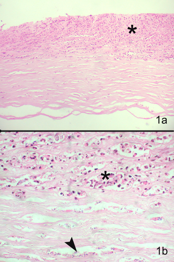
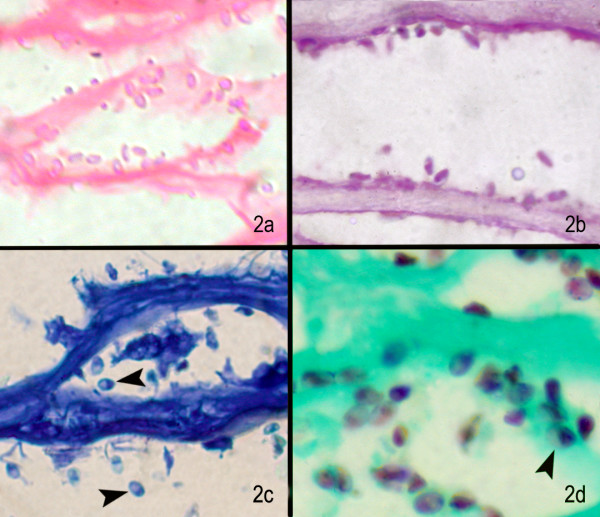
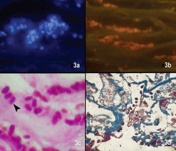
Four cases diagnosed with microsporidiosis on Hematoxylin and Eosin and Periodic Acid Schiff's stained sections of the corneal button between January 2002 and December 2004, were included. Further sections were prospectively stained with calcofluor white, Gram, Giemsa, Masson's trichrome, acridine orange, Gomori's methenamine silver, Gram's chromotrope and modified acid fast stain. The stained sections were analyzed for the spore characteristics in terms of size, shape, color contrast, cell wall morphology, waist band in cytoplasm and ease of detection.

RESULTS

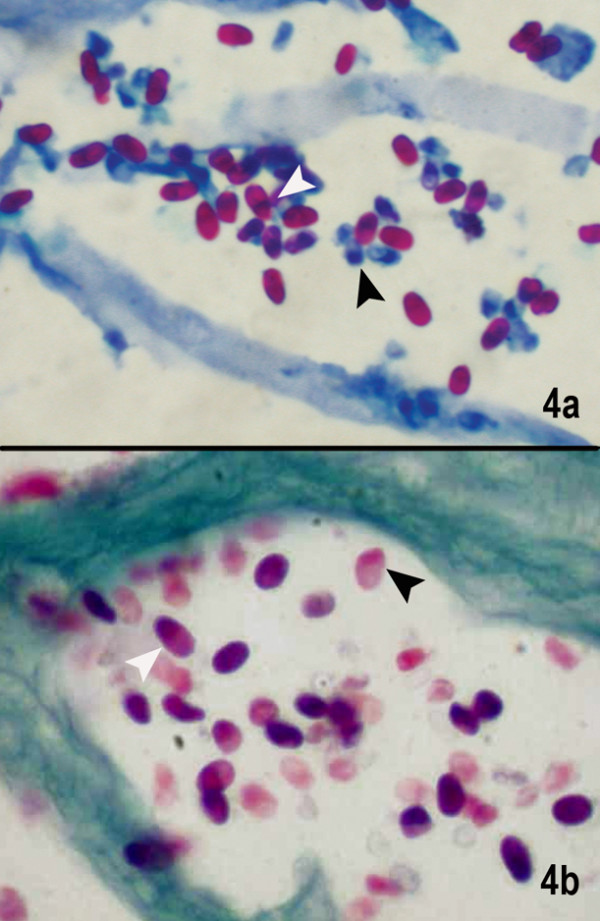
All sections showed microsporidial spores as 3-5 microm, oval bodies. 1% acid fast, Gram's chromotrope and GMS stains provided a reliable diagnosis of microsporidia as diagnostic waist band could be identified and good contrast helped distinguish the spores from inflammatory debris.

CONCLUSION

Considering the ease of performance, cost effectiveness and rapidity of the technique, 1% acid fast stain and Gram's chromotrope stain are ideal for the detection of microsporidia.

摘要

背景

在角膜活检中比较不同染色剂检测微孢子虫的数据有限。因此,我们想评估各种染色剂在角膜组织切片中检测微孢子虫的能力。

方法

纳入2002年1月至2004年12月间经苏木精-伊红染色和过碘酸希夫染色的角膜纽扣标本诊断为微孢子虫病的4例病例。进一步的切片前瞻性地用荧光钙白、革兰氏染色、吉姆萨染色、马森三色染色、吖啶橙、戈莫里六胺银染色、革兰氏铬变素染色和改良抗酸染色。对染色切片的孢子特征进行分析,包括大小、形状、颜色对比、细胞壁形态、细胞质中的腰带以及检测的难易程度。

结果

所有切片均显示微孢子虫孢子为3 - 5微米的椭圆形体。1%抗酸染色、革兰氏铬变素染色和六胺银染色能可靠诊断微孢子虫,因为可识别出诊断性腰带,且良好的对比度有助于将孢子与炎性碎屑区分开来。

结论

考虑到操作的简便性、成本效益和技术的快速性,1%抗酸染色和革兰氏铬变素染色是检测微孢子虫的理想方法。

https://cdn.ncbi.nlm.nih.gov/pmc/blobs/1895/1513581/f9f8d8213db6/1472-6890-6-6-4.jpg
https://cdn.ncbi.nlm.nih.gov/pmc/blobs/1895/1513581/1e46ec633095/1472-6890-6-6-1.jpg
https://cdn.ncbi.nlm.nih.gov/pmc/blobs/1895/1513581/1a012fbf5bfe/1472-6890-6-6-2.jpg
https://cdn.ncbi.nlm.nih.gov/pmc/blobs/1895/1513581/7190b0cc7c4e/1472-6890-6-6-3.jpg
https://cdn.ncbi.nlm.nih.gov/pmc/blobs/1895/1513581/f9f8d8213db6/1472-6890-6-6-4.jpg

相似文献

1
Histopathological evaluation of ocular microsporidiosis by different stains.不同染色法对眼部微孢子虫病的组织病理学评估。
BMC Clin Pathol. 2006 Jun 23;6:6. doi: 10.1186/1472-6890-6-6.
2
Cytologic detection of microsporidia spores in bile. A comparison of stains.胆汁中微孢子虫孢子的细胞学检测:染色方法比较
Acta Cytol. 1999 Mar-Apr;43(2):98-103. doi: 10.1159/000330959.
3
Detection of Microsporidia by different staining techniques.采用不同染色技术检测微孢子虫。
J Egypt Soc Parasitol. 1998 Dec;28(3):729-38.
4
Use of the fluorochrome calcofluor white in the screening of stool specimens for spores of microsporidia.荧光染料白荧光素在粪便标本微孢子虫孢子筛查中的应用。
Am J Clin Pathol. 1995 May;103(5):656-9. doi: 10.1093/ajcp/103.5.656.
5
Clinical and microbiological profile of microsporidial keratoconjunctivitis in southern India.印度南部微孢子虫性角膜结膜炎的临床和微生物学特征
Ophthalmology. 2006 Apr;113(4):531-7. doi: 10.1016/j.ophtha.2005.10.062. Epub 2006 Feb 17.
6
Pragmatic Combination of Available Diagnostic Tools for Optimal Detection of Intestinal Microsporidia.实用组合现有诊断工具以最佳检测肠道微孢子虫。
Adv Exp Med Biol. 2018;1057:85-94. doi: 10.1007/5584_2017_97.
7
Acridine orange as a screen for organisms in clinical specimens and comparison with gram's stain.吖啶橙作为临床标本中微生物的筛选方法及其与革兰氏染色的比较。
Arch Pathol Lab Med. 1988 May;112(5):529-32.
8
Evaluation of hematoxylin and eosin and special stains for the detection of acanthamoeba keratitis in penetrating keratoplasties.苏木精-伊红染色及特殊染色在穿透性角膜移植术中检测棘阿米巴角膜炎的评估
Am J Ophthalmol. 2003 Sep;136(3):520-6. doi: 10.1016/s0002-9394(03)00322-2.
9
A new and improved "quick-hot Gram-chromotrope" technique that differentially stains microsporidian spores in clinical samples, including paraffin-embedded tissue sections.一种新的改良型“快速热染革兰色变剂”技术,可对临床样本(包括石蜡包埋组织切片)中的微孢子虫孢子进行鉴别染色。
Arch Pathol Lab Med. 1997 Aug;121(8):888-93.
10
Fluorescence techniques for diagnosing intestinal microsporidiosis in stool, enteric fluid, and biopsy specimens from acquired immunodeficiency syndrome patients with chronic diarrhea.用于诊断慢性腹泻的获得性免疫缺陷综合征患者粪便、肠液和活检标本中肠道微孢子虫病的荧光技术。
Arch Pathol Lab Med. 1996 Sep;120(9):847-53.

引用本文的文献

1
A Rare Microsporidial Infection in Lamellar Corneal Tissue, following Transepithelial Photorefractive Keratectomy.经上皮光性屈光性角膜切削术后板层角膜组织中的罕见微孢子虫感染
Case Rep Ophthalmol. 2023 Apr 4;14(1):127-133. doi: 10.1159/000528894. eCollection 2023 Jan-Dec.
2
A Novel Three-Dimensional Imaging System Based on Polysaccharide Staining for Accurate Histopathological Diagnosis of Inflammatory Bowel Diseases.基于多糖染色的新型三维成像系统,用于准确的炎症性肠病组织病理学诊断。
Cell Mol Gastroenterol Hepatol. 2022;14(4):905-924. doi: 10.1016/j.jcmgh.2022.07.001. Epub 2022 Jul 12.
3
Comparison of histologic methods for the detection of spores in the gills of Atlantic salmon.

本文引用的文献

1
Clinical and microbiological profile of microsporidial keratoconjunctivitis in southern India.印度南部微孢子虫性角膜结膜炎的临床和微生物学特征
Ophthalmology. 2006 Apr;113(4):531-7. doi: 10.1016/j.ophtha.2005.10.062. Epub 2006 Feb 17.
2
Use of different stains for microscopic evaluation of corneal scrapings for diagnosis of microsporidial keratitis.使用不同染色剂对角膜刮片进行显微镜检查以诊断微孢子虫性角膜炎。
J Clin Microbiol. 2006 Feb;44(2):583-5. doi: 10.1128/JCM.44.2.583-585.2006.
3
Microsporidia of mammals--widespread pathogens or opportunistic curiosities?
大西洋鲑鱼鳃中孢子检测组织学方法的比较
J Vet Diagn Invest. 2020 Jan;32(1):142-146. doi: 10.1177/1040638719887707. Epub 2019 Nov 18.
4
Challenges in the diagnosis of microbial keratitis: A detailed review with update and general guidelines.微生物性角膜炎诊断中的挑战:详细综述及更新与通用指南
Saudi J Ophthalmol. 2019 Jul-Sep;33(3):268-276. doi: 10.1016/j.sjopt.2019.09.002. Epub 2019 Sep 11.
5
A Fatal Case of Disseminated Microsporidiosis Due to in a Renal and Pancreas Allograft Recipient.一例肾和胰腺同种异体移植受者因[未提及具体病因]导致播散性微孢子虫病的致命病例。
Open Forum Infect Dis. 2019 Jul 8;6(7):ofz285. doi: 10.1093/ofid/ofz285. eCollection 2019 Jul.
6
Pharmacokinetics and efficacy of Bioerodible Dexamethasone implant in Concanavalin A-induced uveitic cataract rabbit model.可生物降解地塞米松植入物在伴刀豆球蛋白A诱导的葡萄膜炎性白内障兔模型中的药代动力学及疗效
Pharm Res. 2014 Nov;31(11):3179-90. doi: 10.1007/s11095-014-1410-7. Epub 2014 Jun 20.
7
Unusual fluorescent granulomas and myonecrosis in Danio rerio infected by the microsporidian pathogen Pseudoloma neurophilia.受微孢子虫病原体嗜神经假瘤虫感染的斑马鱼中出现的异常荧光肉芽肿和肌坏死。
Zebrafish. 2014 Jun;11(3):283-90. doi: 10.1089/zeb.2013.0933. Epub 2014 Apr 7.
8
Delayed diagnosis of microsporidial stromal keratitis: unusual Wessely ring presentation and partial treatment with medications against Acanthamoeba.微孢子虫性基质性角膜炎的延迟诊断:不寻常的韦塞利环表现及抗棘阿米巴药物的部分治疗
BMJ Case Rep. 2011 Feb 24;2011:bcr0820103233. doi: 10.1136/bcr.08.2010.3233.
9
Transmission, diagnosis, and recommendations for control of Pseudoloma neurophilia infections in laboratory zebrafish (Danio rerio) facilities.实验室斑马鱼(Danio rerio)设施中嗜神经假瘤菌感染的传播、诊断及控制建议
Comp Med. 2011 Aug;61(4):322-9.
10
Update on pathologic diagnosis of corneal infections and inflammations.角膜感染与炎症的病理诊断进展
Middle East Afr J Ophthalmol. 2011 Oct;18(4):277-84. doi: 10.4103/0974-9233.90128.
哺乳动物的微孢子虫——广泛传播的病原体还是机会性的罕见生物?
Parasitol Today. 1987 Sep;3(9):267-73. doi: 10.1016/0169-4758(87)90103-7.
4
Opportunistic intestinal parasitic infections in human immunodeficiency virus infected patients in Mangalore, South India.印度南部芒格洛尔地区人类免疫缺陷病毒感染患者的机会性肠道寄生虫感染
Trop Doct. 2003 Oct;33(4):250. doi: 10.1177/004947550303300425.
5
Microsporidial keratoconjunctivitis in a HIV-seronegative patient treated with debridement and oral itraconazole.一名接受清创术和口服伊曲康唑治疗的HIV血清阴性患者的微孢子虫性角结膜炎
Am J Ophthalmol. 2003 Oct;136(4):745-6. doi: 10.1016/s0002-9394(03)00391-x.
6
Role of coccidian parasites in causation of diarrhoea in HIV infected patients in Chennai.球虫寄生虫在钦奈HIV感染患者腹泻病因中的作用
Indian J Med Res. 2002 Sep;116:85-9.
7
Microsporidial keratoconjunctivitis in a healthy contact lens wearer without human immunodeficiency virus infection.一名无人类免疫缺陷病毒感染的健康隐形眼镜佩戴者发生微孢子虫性角结膜炎。
Ophthalmology. 2001 May;108(5):976-8. doi: 10.1016/s0161-6420(01)00542-5.
8
Corneal ulceration in the elderly in Hyderabad, south India.印度南部海得拉巴市老年人的角膜溃疡
Br J Ophthalmol. 2000 Jan;84(1):54-9. doi: 10.1136/bjo.84.1.54.
9
Diagnosing Helicobacter pylori by imprint cytology: can the same biopsy specimen be used for histology?通过印片细胞学诊断幽门螺杆菌:同一活检标本能否用于组织学检查?
Diagn Cytopathol. 1998 May;18(5):330-2. doi: 10.1002/(sici)1097-0339(199805)18:5<330::aid-dc4>3.0.co;2-e.
10
Optimal screening and diagnosis of microsporida in tissue sections: a comparison of polarization, special stains, and molecular techniques.组织切片中微孢子虫的最佳筛查与诊断:偏振光、特殊染色及分子技术的比较
Am J Clin Pathol. 1998 Apr;109(4):404-10. doi: 10.1093/ajcp/109.4.404.