Saari Seppo A M, Nikander Sven E
Department of Basic Veterinary Sciences (FINPAR), Faculty of Veterinary Medicine, P,O, Box 66, FIN-00014 University of Helsinki, Finland.
Acta Vet Scand. 2006 Sep 5;48(1):18. doi: 10.1186/1751-0147-48-18.
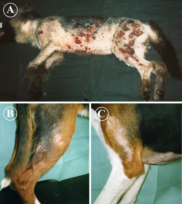
Pelodera (Rhabditis) strongyloides is a small saprophytic nematode that lives in decaying organic matter. On rare occasions, it can invade the mammalian skin, causing a pruritic, erythematous, alopecic and crusting dermatitis on skin sites that come into contact with the ground. Diagnosis of the disease is based on case history (a dog living outdoors on damp straw bedding) with characteristic skin lesions and on the demonstration of typical larvae in skin scrapings or biopsy. Pelodera (rhabditic) dermatitis cases have been reported mainly from Central European countries and the United States.
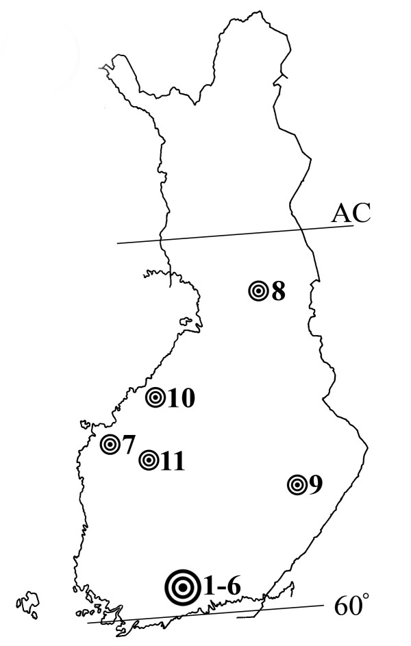
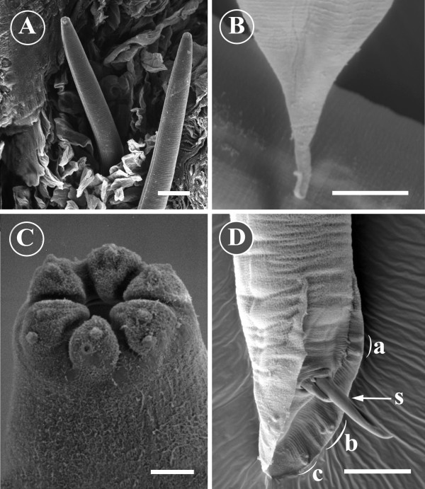
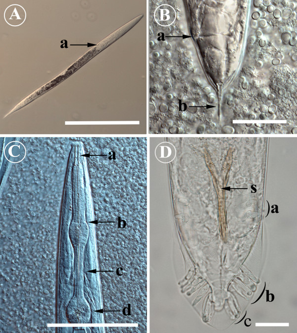
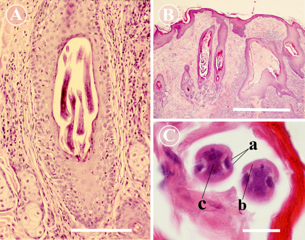
During 1975-1999, we verified 11 canine cases of Pelodera dermatitis in Finland. The cases were confirmed by identifying Pelodera larvae in scrapings. Biopsies for histopathology were obtained from three cases, and typical histopathological lesions (epidermal hyperplasia, epidermal and follicular hyperkeratosis, folliculitis and furunculosis with large numbers of nematode larvae of 25-40 microm of diameter within hair follicles) were present. The Pelodera strongyloides dermatitica strain from the first verified case in Finland has been maintained in ordinary blood agar in our laboratory since 1975. Light microscopy (LM) and scanning electron microscopy (SEM) studies were employed to obtain detailed morphological information about the causative agent. The rhabditiform oesophagus at all developmental stages, the morphology of the anterior end of the nematode, copulatory bursa and spicules of the male and the tail of the female were the most important morphological features for identifying P. strongyloides.
These cases show that Pelodera dermatitis occurs in Finland, and also farther north than described earlier in the literature. This condition should be considered when a dog living outdoors has typical skin lesions situated at sites in contact with the ground as the main presenting clinical feature. The fastest and easiest way to confirm the diagnosis is to demonstrate typical larvae in skin scrapings. In uncertain cases, skin biopsy and culturing of the worms are recommended as supplementary diagnostic procedures.
类圆小杆线虫是一种生活在腐烂有机物中的小型腐生线虫。在极少数情况下,它可侵入哺乳动物皮肤,在与地面接触的皮肤部位引起瘙痒、红斑、脱毛和结痂性皮炎。该病的诊断基于病史(一只生活在户外潮湿稻草垫上的狗)、特征性皮肤病变以及在皮肤刮屑或活检中发现典型幼虫。类圆小杆线虫性皮炎病例主要报道于中欧国家和美国。
1975年至1999年期间,我们在芬兰确诊了11例犬类圆小杆线虫性皮炎病例。通过在刮屑中鉴定出类圆小杆线虫幼虫确诊病例。对3例进行了组织病理学活检,出现了典型的组织病理学病变(表皮增生、表皮和毛囊角化过度、毛囊炎和疖肿,毛囊内有大量直径为25 - 40微米的线虫幼虫)。自1975年以来,芬兰首例确诊病例的类圆小杆线虫皮炎菌株一直在我们实验室的普通血琼脂中保存。采用光学显微镜(LM)和扫描电子显微镜(SEM)研究以获取有关病原体的详细形态学信息。在所有发育阶段的杆状食道、线虫前端的形态、雄性的交合伞和交合刺以及雌性的尾部是鉴定类圆小杆线虫的最重要形态学特征。
这些病例表明类圆小杆线虫性皮炎在芬兰存在,而且比文献中先前描述的更往北。当一只生活在户外的狗以与地面接触部位出现典型皮肤病变为主要临床特征时,应考虑这种情况。确诊的最快且最简单方法是在皮肤刮屑中发现典型幼虫。在不确定的病例中,建议进行皮肤活检和虫体培养作为辅助诊断程序。