Espinola-Zavaleta Nilda, Soto M Elena, Castellanos Luis Muñóz, Játiva-Chávez Silvio, Keirns Candace
Echocardiography in Outpatient Clinic, Instituto Nacional de Cardiología Ignacio Chávez, Juan Badiano No. 1, Colonia Sección XVI Tlalpan, 14080 México, D.F., Mexico.
Cardiovasc Ultrasound. 2006 Sep 26;4:35. doi: 10.1186/1476-7120-4-35.
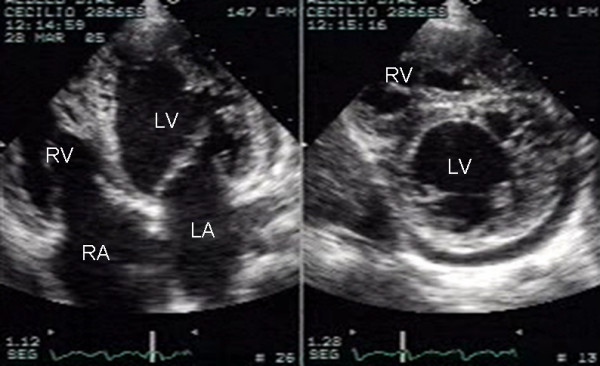
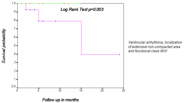
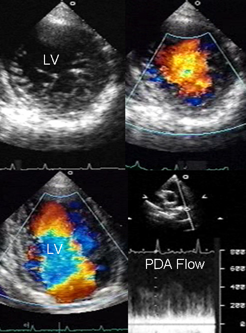
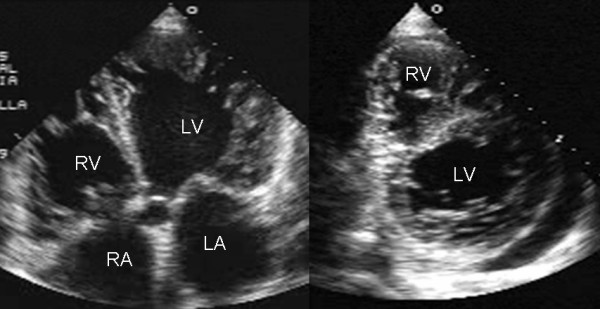
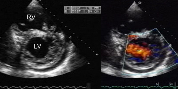
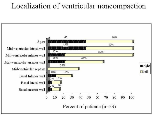
The aim of the present study was to describe the clinical and echocardiographic findings of ventricular noncompaction in adult patients. Fifty-three patients underwent complete clinical history, electrocardiogram, Holter and transthoracic echocardiogram. Forty patients (75%) were in class I/II of the New York Heart Association, and 13 (25%) in class III/IV. Ventricular and supraventricular escape beats were found in 40% and 26.4%, respectively. Holter showed premature ventricular contractions in 32% and sustained ventricular tachycardia in 7.5%. Ventricular noncompaction was an isolated finding in 74% of cases and was associated with other congenital heart disease in 26%. Noncompacted ventricular myocardium involved only left ventricle in 62% of the patients and both ventricles in 38%. The mean ratio of noncompacted to compacted myocardial layers at the site of maximal wall thickness was 3.4 +/- 0.87 mm (range 2.2-7.5). The presence of ventricular noncompaction in more than three segments was associated with a functional class greater than II and ventricular arrhythmia with demonstrable statistical significance by chi2(p < 0.003).
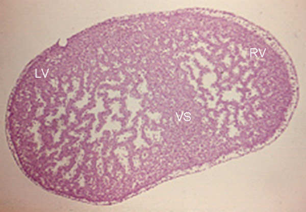
a) Noncompacted cardiomyopathy is a congenital pathological entity that can occur in isolated form or associated with other heart disease and often involves both ventricles. b) A ratio of noncompacted to compacted myocardium greater than 3 and involvement of three or more segments are indicators of poor prognosis. c) Since the clinical manifestations are not sufficient to establish diagnosis, echocardiography is the diagnostic tool that makes it possible to document ventricular noncompaction and establish prognostic factors.
本研究的目的是描述成年患者心室心肌致密化不全的临床和超声心动图表现。53例患者接受了完整的临床病史、心电图、动态心电图和经胸超声心动图检查。40例患者(75%)为纽约心脏协会心功能I/II级,13例(25%)为III/IV级。分别有40%和26.4%的患者出现室性和室上性逸搏。动态心电图显示32%的患者有室性早搏,7.5%的患者有持续性室性心动过速。74%的病例中心室心肌致密化不全是孤立发现,26%与其他先天性心脏病相关。62%的患者非致密化心室心肌仅累及左心室,38%累及双心室。最大室壁厚度处非致密化心肌层与致密化心肌层的平均比值为3.4±0.87mm(范围2.2 - 7.5)。超过三个节段存在心室心肌致密化不全与心功能II级以上及室性心律失常相关,经卡方检验具有显著统计学意义(p < 0.003)。
a)心肌致密化不全性心肌病是一种先天性病理实体,可单独出现或与其他心脏病相关,常累及双心室。b)非致密化心肌与致密化心肌的比值大于3以及三个或更多节段受累是预后不良的指标。c)由于临床表现不足以确诊,超声心动图是能够记录心室心肌致密化不全并确定预后因素的诊断工具。