Lechene Claude, Hillion Francois, McMahon Greg, Benson Douglas, Kleinfeld Alan M, Kampf J Patrick, Distel Daniel, Luyten Yvette, Bonventre Joseph, Hentschel Dirk, Park Kwon Moo, Ito Susumu, Schwartz Martin, Benichou Gilles, Slodzian Georges
National Resource for Imaging Mass Spectrometry, Harvard Medical School and Department of Medicine, Brigham and Women's Hospital, Cambridge, MA 02139, USA.
J Biol. 2006;5(6):20. doi: 10.1186/jbiol42.
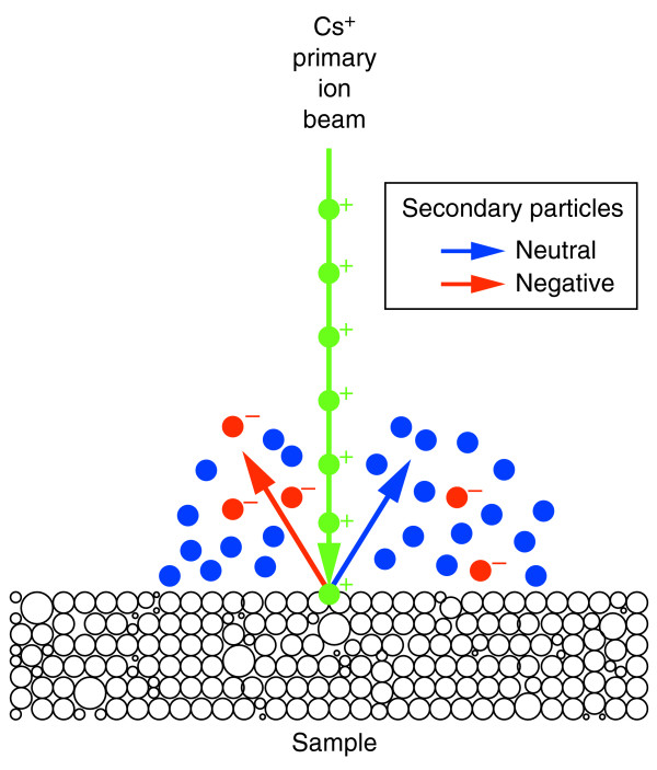
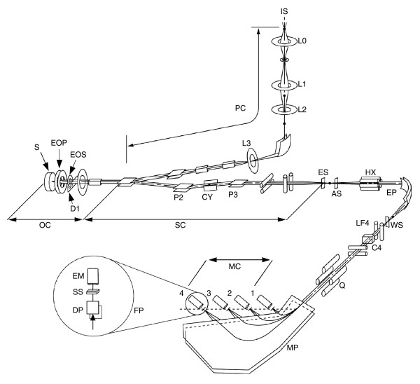
Secondary-ion mass spectrometry (SIMS) is an important tool for investigating isotopic composition in the chemical and materials sciences, but its use in biology has been limited by technical considerations. Multi-isotope imaging mass spectrometry (MIMS), which combines a new generation of SIMS instrument with sophisticated ion optics, labeling with stable isotopes, and quantitative image-analysis software, was developed to study biological materials.
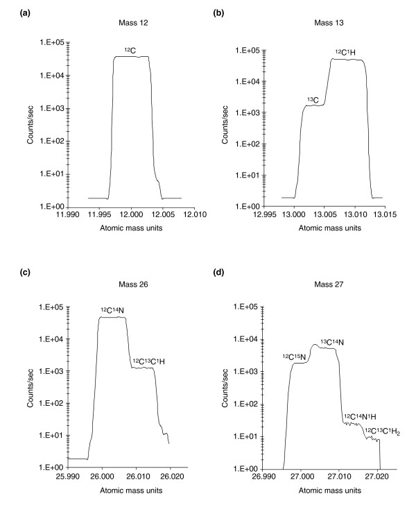
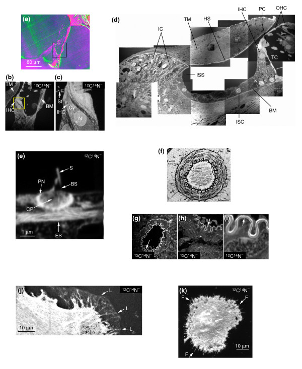
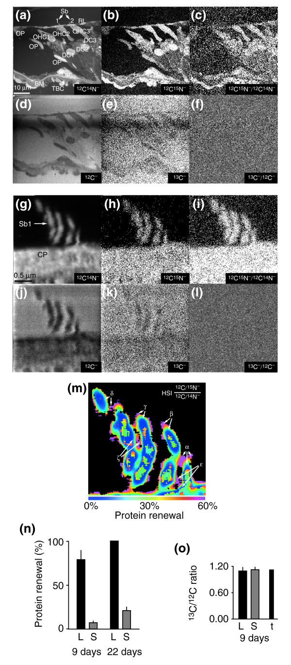
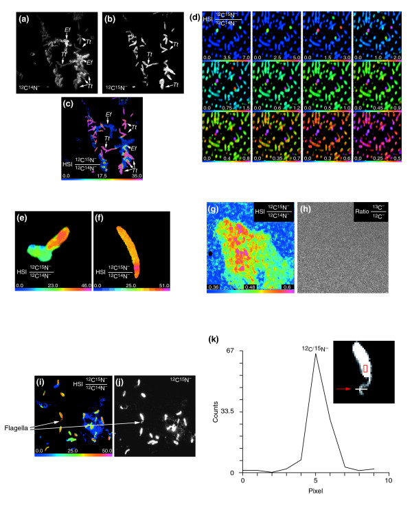
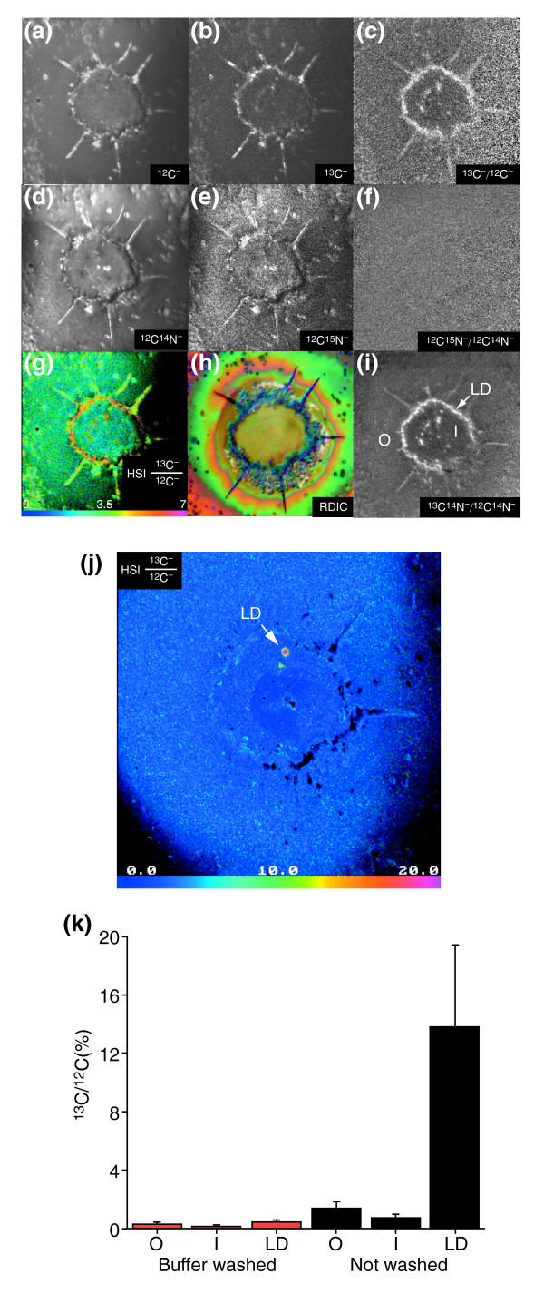
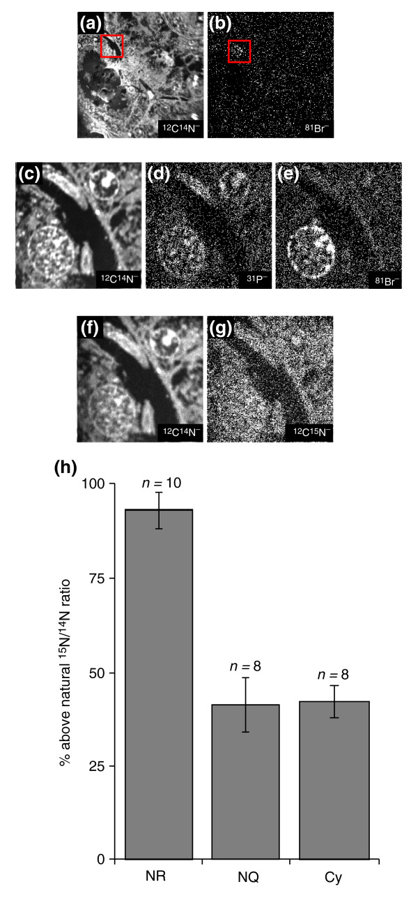
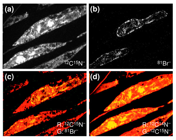
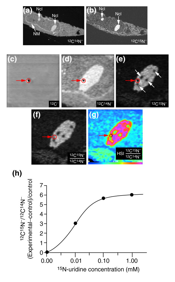
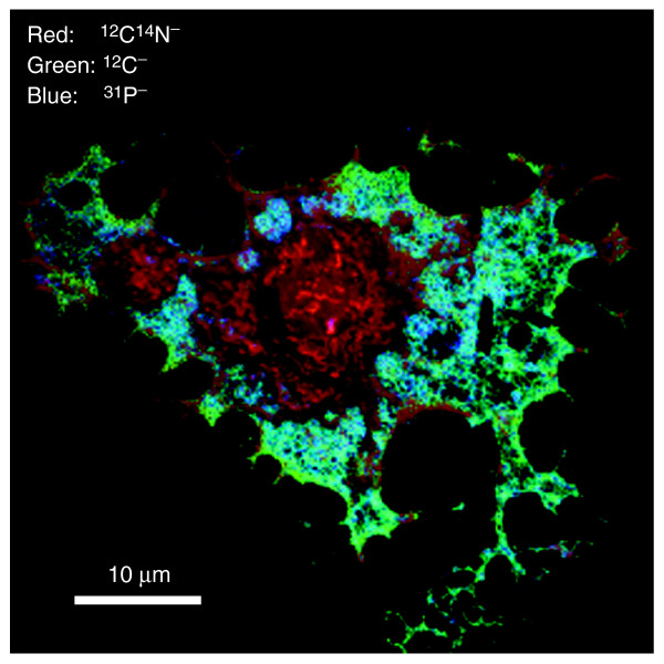
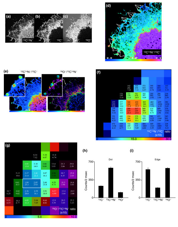
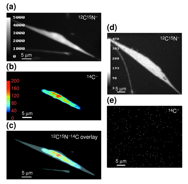
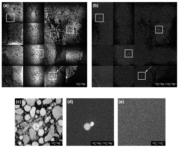
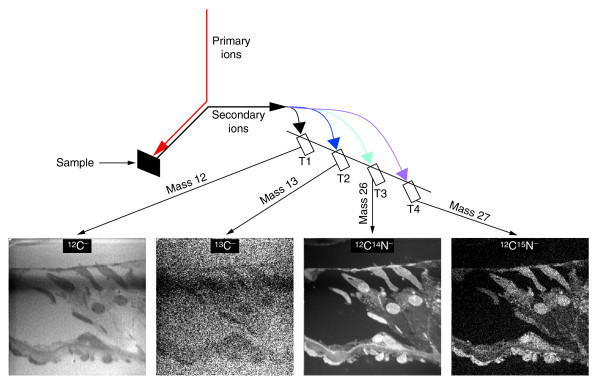
The new instrument allows the production of mass images of high lateral resolution (down to 33 nm), as well as the counting or imaging of several isotopes simultaneously. As MIMS can distinguish between ions of very similar mass, such as 12C15N- and 13C14N-, it enables the precise and reproducible measurement of isotope ratios, and thus of the levels of enrichment in specific isotopic labels, within volumes of less than a cubic micrometer. The sensitivity of MIMS is at least 1,000 times that of 14C autoradiography. The depth resolution can be smaller than 1 nm because only a few atomic layers are needed to create an atomic mass image. We illustrate the use of MIMS to image unlabeled mammalian cultured cells and tissue sections; to analyze fatty-acid transport in adipocyte lipid droplets using 13C-oleic acid; to examine nitrogen fixation in bacteria using 15N gaseous nitrogen; to measure levels of protein renewal in the cochlea and in post-ischemic kidney cells using 15N-leucine; to study DNA and RNA co-distribution and uridine incorporation in the nucleolus using 15N-uridine and 81Br of bromodeoxyuridine or 14C-thymidine; to reveal domains in cultured endothelial cells using the native isotopes 12C, 16O, 14N and 31P; and to track a few 15N-labeled donor spleen cells in the lymph nodes of the host mouse.
MIMS makes it possible for the first time to both image and quantify molecules labeled with stable or radioactive isotopes within subcellular compartments.
二次离子质谱(SIMS)是化学和材料科学中研究同位素组成的重要工具,但其在生物学中的应用一直受到技术因素的限制。多同位素成像质谱(MIMS)结合了新一代SIMS仪器、精密离子光学、稳定同位素标记和定量图像分析软件,用于研究生物材料。
新仪器能够生成高横向分辨率(低至33纳米)的质量图像,同时对几种同位素进行计数或成像。由于MIMS能够区分质量非常相似的离子,如12C15N-和13C14N-,它能够在小于一立方微米的体积内精确且可重复地测量同位素比率,从而测量特定同位素标记的富集水平。MIMS的灵敏度至少是14C放射自显影的1000倍。深度分辨率可小于1纳米,因为只需几个原子层就能创建原子质量图像。我们展示了MIMS用于对未标记的哺乳动物培养细胞和组织切片进行成像;使用13C-油酸分析脂肪细胞脂滴中的脂肪酸转运;使用15N气态氮检测细菌中的固氮作用;使用15N-亮氨酸测量耳蜗和缺血后肾细胞中的蛋白质更新水平;使用15N-尿苷、溴脱氧尿苷的81Br或14C-胸腺嘧啶研究核仁中的DNA和RNA共分布以及尿苷掺入;使用天然同位素12C、16O、14N和31P揭示培养内皮细胞中的结构域;以及追踪宿主小鼠淋巴结中少数15N标记的供体脾细胞。
MIMS首次使在亚细胞区室中对用稳定或放射性同位素标记的分子进行成像和定量成为可能。