Granziera Cristina, DaSilva Alexandre F M, Snyder Josh, Tuch David S, Hadjikhani Nouchine
Athinoula A. Martinos Center for Biomedical Imaging, Massachusetts General Hospital, Harvard Medical School, Charlestown, Massachusetts, United States of America.
PLoS Med. 2006 Oct;3(10):e402. doi: 10.1371/journal.pmed.0030402.
Patients suffering from migraine with aura (MWA) and migraine without aura (MWoA) show abnormalities in visual motion perception during and between attacks. Whether this represents the consequences of structural changes in motion-processing networks in migraineurs is unknown. Moreover, the diagnosis of migraine relies on patient's history, and finding differences in the brain of migraineurs might help to contribute to basic research aimed at better understanding the pathophysiology of migraine.
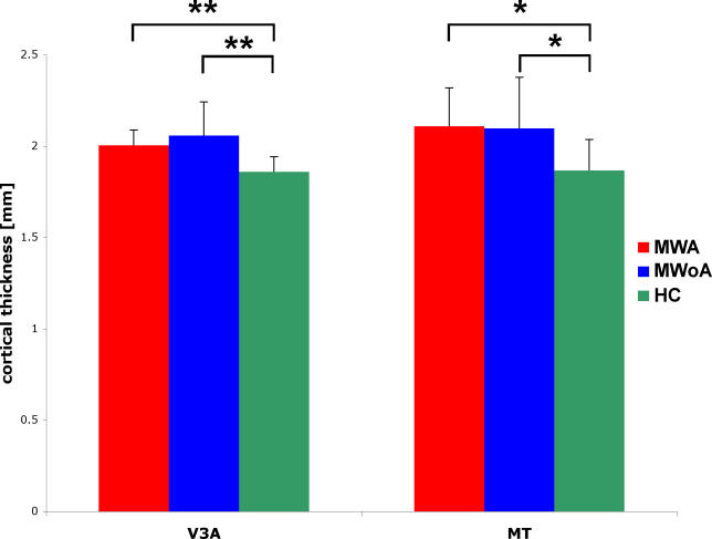
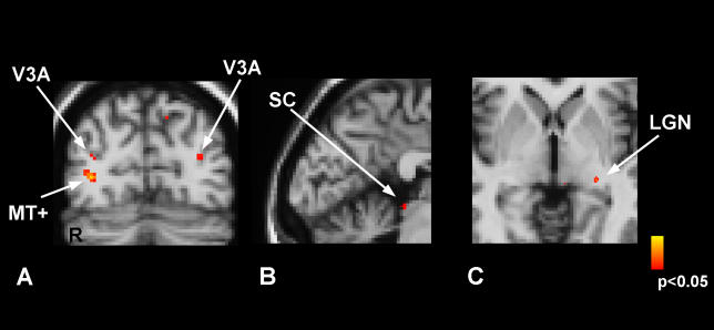
To investigate a common potential anatomical basis for these disturbances, we used high-resolution cortical thickness measurement and diffusion tensor imaging (DTI) to examine the motion-processing network in 24 migraine patients (12 with MWA and 12 MWoA) and 15 age-matched healthy controls (HCs). We found increased cortical thickness of motion-processing visual areas MT+ and V3A in migraineurs compared to HCs. Cortical thickness increases were accompanied by abnormalities of the subjacent white matter. In addition, DTI revealed that migraineurs have alterations in superior colliculus and the lateral geniculate nucleus, which are also involved in visual processing.
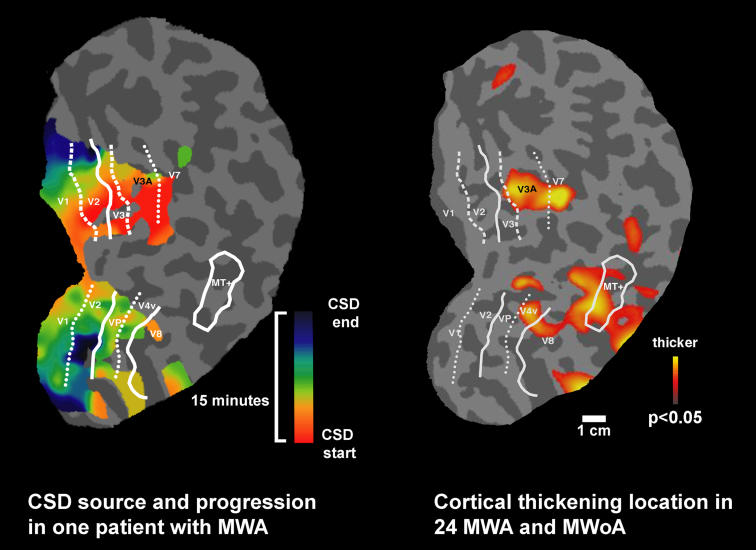
A structural abnormality in the network of motion-processing areas could account for, or be the result of, the cortical hyperexcitability observed in migraineurs. The finding in patients with both MWA and MWoA of thickness abnormalities in area V3A, previously described as a source in spreading changes involved in visual aura, raises the question as to whether a "silent" cortical spreading depression develops as well in MWoA. In addition, these experimental data may provide clinicians and researchers with a noninvasively acquirable migraine biomarker.
伴有先兆的偏头痛(MWA)和无先兆偏头痛(MWoA)患者在发作期间及发作间歇期的视觉运动感知存在异常。这是否代表偏头痛患者运动处理网络结构变化的后果尚不清楚。此外,偏头痛的诊断依赖于患者病史,而在偏头痛患者大脑中发现差异可能有助于推动旨在更好理解偏头痛病理生理学的基础研究。
为了探究这些紊乱的共同潜在解剖学基础,我们使用高分辨率皮质厚度测量和扩散张量成像(DTI)来检查24名偏头痛患者(12名MWA患者和12名MWoA患者)以及15名年龄匹配的健康对照者(HCs)的运动处理网络。我们发现,与HCs相比,偏头痛患者运动处理视觉区域MT+和V3A的皮质厚度增加。皮质厚度增加伴随着其下方白质的异常。此外,DTI显示偏头痛患者上丘和外侧膝状体也有改变,这些结构也参与视觉处理。
运动处理区域网络的结构异常可能是偏头痛患者观察到的皮质兴奋性过高的原因或结果。在MWA和MWoA患者中均发现V3A区域厚度异常,该区域先前被描述为视觉先兆中扩散性变化的源头,这就提出了一个问题,即MWoA中是否也会出现“沉默”的皮质扩散性抑制。此外,这些实验数据可能为临床医生和研究人员提供一种可通过非侵入性获得的偏头痛生物标志物。