• 文献检索
  • 文档翻译
  • 深度研究
  • 学术资讯
  • Suppr Zotero 插件Zotero 插件
  • 邀请有礼
  • 套餐&价格
  • 历史记录
应用&插件
Suppr Zotero 插件Zotero 插件浏览器插件Mac 客户端Windows 客户端微信小程序
定价
高级版会员购买积分包购买API积分包
服务
文献检索文档翻译深度研究API 文档MCP 服务
关于我们
关于 Suppr公司介绍联系我们用户协议隐私条款
关注我们

Suppr 超能文献

核心技术专利:CN118964589B侵权必究
粤ICP备2023148730 号-1Suppr @ 2026

文献检索

告别复杂PubMed语法,用中文像聊天一样搜索,搜遍4000万医学文献。AI智能推荐,让科研检索更轻松。

立即免费搜索

文件翻译

保留排版,准确专业,支持PDF/Word/PPT等文件格式,支持 12+语言互译。

免费翻译文档

深度研究

AI帮你快速写综述,25分钟生成高质量综述,智能提取关键信息,辅助科研写作。

立即免费体验

超声心动图与电子束计算机断层扫描评估瓣膜性主动脉狭窄患者主动脉瓣面积的比较。

A comparison of echocardiographic and electron beam computed tomographic assessment of aortic valve area in patients with valvular aortic stenosis.

作者信息

Piers Lieuwe H, Dikkers Riksta, Tio René A, van den Berg Maarten P, Willems Tineke P, Zijlstra Felix, Oudkerk Matthijs

机构信息

Department of Cardiology, University Medical Centre Groningen, University of Groningen, Hanzeplein 1, P.O. Box 30001, Groningen, 9700 RB, The Netherlands.

出版信息

Int J Cardiovasc Imaging. 2007 Dec;23(6):781-8. doi: 10.1007/s10554-007-9208-x. Epub 2007 Feb 7.

DOI:10.1007/s10554-007-9208-x
PMID:17285263
原文链接:https://pmc.ncbi.nlm.nih.gov/articles/PMC2048829/
Abstract

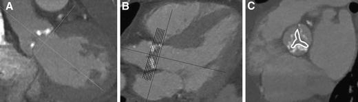
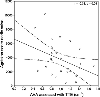
The purpose of this study was to compare electron beam computed tomography (EBT) with transthoracic echocardiography (TTE) in determining aortic valve area (AVA). Thirty patients (9 females, 21 males) underwent a contrast-enhanced EBT scan (e-Speed, GE, San Francisco, CA, USA) and TTE within 17 +/- 12 days. In end-inspiratory breath hold, a prospectively ecg-triggered scan was acquired with a beam speed of 50-100 ms, a collimation of 2 x 1.5 mm and an increment of 3.0 mm. The AVA was measured with planimetry. A complete TTE study was performed in all patients, and the AVA was computed using the continuity equation. There was close correlation between AVA measured with EBT and AVA assessed with TTE (r = 0.60, P < 0.01). The AVA measured with EBT was 0.51 +/- 0.46 cm(2 )larger than the AVA calculated with TTE measurements. EBT appeared to be a valuable non-invasive method to measure the AVA. EBT measures the anatomical AVA, while with TTE the functional AVA is calculated, which explains the difference in results between the methods.

摘要

本研究的目的是比较电子束计算机断层扫描(EBT)与经胸超声心动图(TTE)在测定主动脉瓣面积(AVA)方面的差异。30例患者(9例女性,21例男性)在17±12天内接受了对比增强EBT扫描(e-Speed,GE,美国加利福尼亚州旧金山)和TTE检查。在吸气末屏气状态下,采用前瞻性心电图触发扫描,束流速度为50 - 100毫秒,准直为2×1.5毫米,增量为3.0毫米。通过平面测量法测量AVA。所有患者均进行了完整的TTE检查,并使用连续性方程计算AVA。EBT测量的AVA与TTE评估的AVA之间存在密切相关性(r = 0.60,P < 0.01)。EBT测量的AVA比TTE测量计算出的AVA大0.51±0.46平方厘米。EBT似乎是一种测量AVA的有价值的非侵入性方法。EBT测量的是解剖学上的AVA,而TTE计算的是功能性AVA,这解释了两种方法结果的差异。

https://cdn.ncbi.nlm.nih.gov/pmc/blobs/20d2/2048829/4af817195da6/10554_2007_9208_Fig3_HTML.jpg
https://cdn.ncbi.nlm.nih.gov/pmc/blobs/20d2/2048829/bda449f04907/10554_2007_9208_Fig1_HTML.jpg
https://cdn.ncbi.nlm.nih.gov/pmc/blobs/20d2/2048829/fa0b4c919436/10554_2007_9208_Fig2_HTML.jpg
https://cdn.ncbi.nlm.nih.gov/pmc/blobs/20d2/2048829/4af817195da6/10554_2007_9208_Fig3_HTML.jpg
https://cdn.ncbi.nlm.nih.gov/pmc/blobs/20d2/2048829/bda449f04907/10554_2007_9208_Fig1_HTML.jpg
https://cdn.ncbi.nlm.nih.gov/pmc/blobs/20d2/2048829/fa0b4c919436/10554_2007_9208_Fig2_HTML.jpg
https://cdn.ncbi.nlm.nih.gov/pmc/blobs/20d2/2048829/4af817195da6/10554_2007_9208_Fig3_HTML.jpg

相似文献

1
A comparison of echocardiographic and electron beam computed tomographic assessment of aortic valve area in patients with valvular aortic stenosis.超声心动图与电子束计算机断层扫描评估瓣膜性主动脉狭窄患者主动脉瓣面积的比较。
Int J Cardiovasc Imaging. 2007 Dec;23(6):781-8. doi: 10.1007/s10554-007-9208-x. Epub 2007 Feb 7.
2
Comparison of dual-source computed tomography for the quantification of the aortic valve area in patients with aortic stenosis versus transthoracic echocardiography and invasive hemodynamic assessment.双源 CT 对主动脉瓣狭窄患者主动脉瓣口面积的定量评估与经胸超声心动图及有创血流动力学评估的比较。
Am J Cardiol. 2009 Dec 1;104(11):1561-7. doi: 10.1016/j.amjcard.2009.07.024.
3
Aortic valve area assessment: multidetector CT compared with cine MR imaging and transthoracic and transesophageal echocardiography.主动脉瓣面积评估:多层螺旋CT与电影磁共振成像及经胸和经食管超声心动图的比较
Radiology. 2007 Sep;244(3):745-54. doi: 10.1148/radiol.2443061127. Epub 2007 Jul 13.
4
Differences in aortic valve area measured with CT planimetry and echocardiography (continuity equation) are related to divergent estimates of left ventricular outflow tract area.通过CT平面测量法和超声心动图(连续方程)测量的主动脉瓣面积差异与左心室流出道面积的不同估计值相关。
AJR Am J Roentgenol. 2009 Jun;192(6):1668-73. doi: 10.2214/AJR.08.1986.
5
Effect of the ellipsoid shape of the left ventricular outflow tract on the echocardiographic assessment of aortic valve area in aortic stenosis.左心室流出道椭圆形形态对主动脉瓣狭窄时主动脉瓣面积超声心动图评估的影响。
J Cardiovasc Comput Tomogr. 2014 Jan-Feb;8(1):52-7. doi: 10.1016/j.jcct.2013.12.006. Epub 2014 Jan 11.
6
Echocardiographic and electron beam tomographic assessment of stenosis in patients with aortic valve disease: gradient versus valve area.主动脉瓣疾病患者狭窄的超声心动图和电子束断层扫描评估:压力阶差与瓣膜面积
Neth Heart J. 2006 Oct;14(10):325-329.
7
Three-dimensional imaging of the left ventricular outflow tract: impact on aortic valve area estimation by the continuity equation.左心室流出道的三维成像:连续方程对主动脉瓣口面积估测的影响。
J Am Soc Echocardiogr. 2012 Jul;25(7):749-57. doi: 10.1016/j.echo.2012.05.001. Epub 2012 May 19.
8
Cardiovascular magnetic resonance imaging and transthoracic echocardiography in the assessment of stenotic aortic valve area: a comparative study.心血管磁共振成像与经胸超声心动图在评估主动脉瓣狭窄面积中的比较研究
Acta Radiol. 2012 Nov 1;53(9):995-1003. doi: 10.1258/ar.2012.120261. Epub 2012 Sep 14.
9
Aortic stenosis: comparative evaluation of 16-detector row CT and echocardiography.主动脉瓣狭窄:16排CT与超声心动图的对比评估
Radiology. 2006 Jul;240(1):47-55. doi: 10.1148/radiol.2393050458. Epub 2006 May 18.
10
Aortic valve area assessed with 320-detector computed tomography: comparison with transthoracic echocardiography.320 排 CT 评估主动脉瓣口面积:与经胸超声心动图的比较。
Int J Cardiovasc Imaging. 2014 Jan;30(1):165-73. doi: 10.1007/s10554-013-0295-6. Epub 2013 Oct 15.

本文引用的文献

1
Absolute assessment of aortic valve stenosis by planimetry using cardiovascular magnetic resonance imaging: comparison with transesophageal echocardiography, transthoracic echocardiography, and cardiac catheterisation.使用心血管磁共振成像通过平面测量法对主动脉瓣狭窄进行绝对评估:与经食管超声心动图、经胸超声心动图及心导管检查的比较
Eur J Radiol. 2006 Aug;59(2):276-83. doi: 10.1016/j.ejrad.2006.02.011.
2
Aortic stenosis: comparative evaluation of 16-detector row CT and echocardiography.主动脉瓣狭窄:16排CT与超声心动图的对比评估
Radiology. 2006 Jul;240(1):47-55. doi: 10.1148/radiol.2393050458. Epub 2006 May 18.
3
CT of valvular heart disease.
心脏瓣膜病的CT检查
Int J Cardiovasc Imaging. 2005 Feb;21(1):105-13. doi: 10.1007/s10554-004-5339-5.
4
Evaluation and clinical implications of aortic valve calcification measured by electron-beam computed tomography.电子束计算机断层扫描测量主动脉瓣钙化的评估及临床意义
Circulation. 2004 Jul 20;110(3):356-62. doi: 10.1161/01.CIR.0000135469.82545.D0. Epub 2004 Jul 12.
5
Crossing the aortic valve in severe aortic stenosis: no longer acceptable?在严重主动脉瓣狭窄中穿过主动脉瓣:不再被接受了吗?
J Heart Valve Dis. 2004 May;13(3):344-6.
6
Quantification of aortic valve calcification with electron beam tomography: a histomorphometric validation study.电子束断层扫描对主动脉瓣钙化的定量分析:一项组织形态计量学验证研究。
Invest Radiol. 2004 Apr;39(4):230-4. doi: 10.1097/01.rli.0000115749.08234.6a.
7
Hydraulic formula for calculation of the area of the stenotic mitral valve, other cardiac valves, and central circulatory shunts. I.用于计算二尖瓣狭窄、其他心脏瓣膜及中心循环分流面积的水力公式。I.
Am Heart J. 1951 Jan;41(1):1-29. doi: 10.1016/0002-8703(51)90002-6.
8
Usefulness of aortic valve calcium scores by electron beam computed tomography as a marker for aortic stenosis.电子束计算机断层扫描测定的主动脉瓣钙化积分作为主动脉瓣狭窄标志物的实用性。
Am J Cardiol. 2003 Aug 1;92(3):349-53. doi: 10.1016/s0002-9149(03)00646-5.
9
Reproducibility of electron-beam CT measures of aortic valve calcification.电子束CT测量主动脉瓣钙化的可重复性。
Acad Radiol. 2002 Oct;9(10):1122-7. doi: 10.1016/s1076-6332(03)80513-5.
10
Correlation of degree of aortic valve stenosis by Doppler echocardiogram to quantity of calcium in the valve by electron beam tomography.多普勒超声心动图测量的主动脉瓣狭窄程度与电子束断层扫描测量的瓣膜钙含量的相关性。
Am J Cardiol. 2002 Sep 1;90(5):554-7. doi: 10.1016/s0002-9149(02)02537-7.